|
EnzyChromTM Fructose Assay Kit (EFRU-100)
Quantitative Colorimetric Fructose Determination at
565nm
DESCRIPTION
FRUCTOSE (C6H12O6, also called levulose or
laevulose), is a monosaccharide found in honey, tree fruits, berries,
melons, and some root vegetables along with glucose and galactose. The
human body can use fructose for energy, however, too much consumption
may lead to high triglycerides. Simple, direct and high-throughput
assays for fructose determination find wide applications. BioAssay
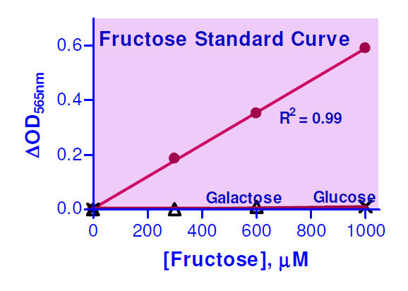
Systems' reagent systems reacts directly and specifically with fructose
to form a colored product. Glucose and galactose do not interfere. The
color intensity at 565nm is directly proportional to the fructose
concentration in the sample.
KEY FEATURES
Use as little as 20 μL samples. Linear detection
range in 96-well plate: 12 to 1000 μM fructose.
APPLICATIONS
Direct Assays:
fructose in biological
samples (e.g. serum, plasma, urine, saliva, milk, culture medium), food,
juice, beverage and other agricultural products.
Drug Discovery/Pharmacology: effects of drugs on
fructose metabolism.
KIT CONTENTS
Assay Buffer:
10 mL Enzyme: 120 μL
PMS Solution: 1.5 mL MTT Solution: 1.5 mL
Standard: 400 μL 20 mM D-Fructose
Storage conditions. The kit is shipped on ice.
Store all components at -20°C. Shelf life of three months after receipt.
Precautions: reagents are for research use only.
Normal precautions for laboratory reagents should be exercised while
using the reagents. Please refer to Material Safety Data Sheet for
detailed information.
ASSAY PROCEDURE
Note: (1) The following substances interfere and
should be avoided in sample preparation: ascorbic acid, SDS (>0.2%),
sodium azide, NP-40 (>1%) and Tween-20 (>1%). (2) This assay is based on
a kinetic reaction. To ensure identical incubation time, addition of
Working Reagent to standard and samples should be quick and mixing
should be brief but thorough. Use of a multi-channel pipettor is
recommended.
Sample treatment: liquid samples such as serum,
plasma and fruit juices can be assayed directly. Because fruit juices
may contain high concentrations of fructose, it is recommended to dilute
juice sample 50- fold (n = 50) in dH2O prior to assay. Milk
samples should be cleared by mixing 600 μL milk with 100 μL 6 N HCl.
Centrifuge 5 min at 14,000 rpm. Transfer 300 μL supernatant into a clean
tube and neutralize with 50 μL 6 N NaOH. The neutralized supernatant is
ready for assay (dilution factor n = 1.36).
1. Equilibrate all components to room temperature.
Briefly centrifuge the tubes before opening. Keep thawed tubes on ice
during assay.
2.
Standards: mix 12 μL 20 mM Standard with 228 μL dH2O (final 1000
μM). Dilute standard in dH2O as follows.
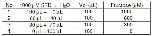
Transfer 20 μL diluted standards into separate wells
of a clear flatbottom 96-well plate.
Samples: transfer 20 μL of each sample into
separate wells of the plate.
3. Color reaction. Prepare enough Working
Reagent by mixing, for each reaction well, 56 μL Assay Buffer, 1 μL
Enzyme, 14 μL PMS Solution and 14 μL MTT Solution. Keep Working Reagent
protected from light. Add 80 μL Working Reagent to each well. Tap plate
to mix. Do not
expose Working
Reagent to light for more than 5 minutes.
Incubate 60 min at room
temperature in the dark.
4. Read optical density at 565nm (520-600nm).
Note:
If the calculated fructose concentration of a sample is higher than 1000
μM, dilute sample in water and repeat the assay. Multiply result by the
dilution factor n.
CALCULATION
Subtract blank value (water, #4) from the standard
values and plot the DOD against standard concentrations. Determine the
slope and calculate the fructose concentration of Sample,
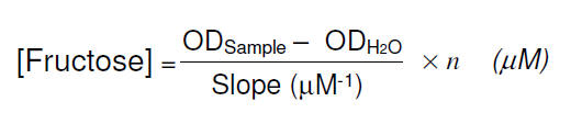
ODSAMPLE, ODH2O are optical density values
of the sample and water. n is the dilution factor.
Conversions: 1 mM fructose equals 18 mg/dL, 0.018%
or 180 ppm.
MATERIALS REQUIRED, BUT NOT PROVIDED
Pipetting devices, centrifuge tubes, clear flat-bottom
uncoated 96-well plates, optical density plate reader.
LITERATURE
1. Novelli G, Reichardt JK. (2000). Molecular basis of
disorders of human fructose metabolism: past, present, and future. Mol
Genet Metab. 71:62-65.
2. Pudek MR et al. (1990). Low concentration fructose
determination in plasma adapted to the Cobas-Bio. Clin Biochem.
23:221-223.
3. Gabrielli M. (1978). Serum fructose determination
with centrifugal analyzers. Clin. Chem. 24:1990-1995. |


