|


|
OUR SUPPLIERS
KOMABIOTECH
301,
Gayang Technotown, #1487
Gayang 3 dong,
Gangseo-gu
Seoul 157-793, KOREA
Spherotech, Inc.
27845 Irma Lee Circle, Unit 101
Lake Forest, IL 60045
|
|
Exalpha
Biologicals,
Inc.
2 Shaker Road,
Unit B101
Shirley, MA
01464
SCETI K.K
BIOSCIENCE Export DF Kasumigaseki Place,3-6-7 Kasumigaseki Chiyoda-ku, Tokyo 100-0013 JAPAN
|
EY
Laboratories, Inc. Headquarters
107 N.
Amphlett Blvd
San Mateo, CA. 94401 USA
EXBIO Praha, a.s.
Nad
Safinou II 366
252 42 Vestec
Czech Republic
Sacace Biotechnologies S.r.l.
Via Scalabrini,
44
22100 Como Italy
 GENTAUR BVBA
VAT BE0473327336
Av. de l Armee 68 B4
1040 Brussels
BELGIUM
Tel + 32 16 58 90
45
Fax + 32 16 50 90 45
 GENTAUR France SARL
SIRET 48423788800017
Rue Lagrange, 9
75005 Paris,
France
Tel 01 43 25 01 50
Fax 01 43 25 01 60
 GENTAUR Germany
Marienbongard 20
GENTAUR Germany
Marienbongard 20
52074 Aachen,
Germany
Tel 0241 56 00
99 68
Fax 0241 56 00 47 88
 GENTAUR
Pol Sp. Z.o.o. Ulica
Ogarna 15/19B m2 GENTAUR
Pol Sp. Z.o.o. Ulica
Ogarna 15/19B m2
80-826 GDANSK
Tel 00 48 58 760 77
08
Fax: 00 32 16 50 90
45
 GENTAUR Italy
23015 Milano, Italy
Tel 02 36 00 65
93
Fax 02 36 00 65
94
 Česká republika
Praha
Česká republika
Praha
+420246019719
 Danmark
+4569918806
 Finland Helsset
+358942419041
 Ελλάς Αθήνα
+302111768494
 Ireland Dublin
+35316526556
 Luxembourg
+35220880274
 Magyarország
Budapest
Magyarország
Budapest
+3619980547
 Nederland
+31208080893
 Norge
Oslo Norge
Oslo
+4721031366
 Österreich
+43720880899
 Sverige
Stockholm Sverige
Stockholm
+46852503438
 Schweiz Züri
+41435006251
Northern America
 Canada Montreal
+15149077481
 US New York
+17185132983
Other Countries
0032 (0)16 41 44 07 |
|
|
|
|
|
|

| |
This Package
Insert is provided for product evaluation purposes only and is
not intended to be used in place of the Package Insert shipped with the
product.
IMMUNO-TEK
Quantitative
Human IgG Antigen ELISA
FOR RESEARCH
USE ONLY.
NOT FOR in vitro DIAGNOSTIC USE.
|
|
|
|
|
INTENDED
USE |
|
The Immuno-Tek Human IgG EIA Kit is a rapid, easy to use enzyme
linked immunosorbant assay (EIA) designed for the measurement of
human IgG in cell culture supernatants, ascites or other
biological fluid. The kit is especially useful in monitoring the
production and purification of mouse monoclonal antibodies. The
kit contains premixed reagents and takes less than two hours to
obtain results. The microplate and detector antibody in the kit
have been specifically balanced to react uniformly with all
subclasses of human IgG.
The Immuno-Tek Human IgG EIA Kit is for Research Purposes Only.
|
|
|
|
|
|
PRINCIPLE OF
THE TEST |
|
Microwells coated with polyclonal antibodies to human IgG form
the capture phase of the assay. These antibodies bind uniformly
to all subclasses of human IgG. Captured human IgG then reacts
with detector antibody which is a polyclonal anti-human IgG
conjugated with horseradish peroxidase. This reagent also reacts
uniformly to all subclasses of human IgG. Enzyme activity in the
wells are then quantified using tetramethyl benzidine as a
substrate. |
|
|
|
|
|
REAGENTS |
|
Materials Supplied
-
Microplate,
(1x96 well):
Strips coated with purified Goat Anti-Human IgG
-
Detector
Antibody (12 ml):
Contains
conjugated Goat Anti-Human IgG peroxidase
-
Human IgG
Standard (5 ml):
Contains Human
IgG and assay diluent
-
Assay Diluent
(100 ml):
Contains PBS,
Triton X-100(R)
and 2-Chloroacetamide
-
Plate Wash
Buffer (125 ml):
Contains PBS,
Tween 20(R)
and 2-Chloroacetamide
-
Substrate (12
ml):
Contains Tetramethyl Benzidine (TMB)
-
Stop Solution
(12 ml):
Contains 2M Sulfuric Acid
-
Microtiter
Plate Sealers (1 pk):
10 sealers per pack
-
Plastic Bag (1
bag):
For storage of unused microtiter plate strips
(R)
Triton X-100 is a registered trademark of Rohm and Haas. Tween
20 is a registered trademark of Imperial Chemical Industries.
Storage
Store all kit
reagents at 2-8 ー
C. Do not freeze.
Materials
Required but not Supplied
-
Test tubes and
racks for preparing specimen and IgG standard dilutions
-
Adjustable
micropipets, single and multi-channel
-
Distilled or
deionized water
-
Incubator
capable of maintaining 37 + 1ー
C
-
Graduated
cylinders and assorted beakers
-
Automatic
microtiter plate washer or manual vacuum aspiration
equipment
|
|
|
|
|
|
PRECAUTIONS |
|
FOR RESEARCH
USE ONLY. Not For in vitro Diagnostic Use. |
|
|
|
|
|
PREPARATION
OF REAGENTS |
|
Plate Wash Buffer
Dilute 10X
Plate Wash Buffer 1:10 in distilled or deionized water prior to
use. Mix thoroughly. Prepared 1X Plate Wash Buffer can be stored
at 2-8ー C
for up to one week. Additional 10X Plate Wash Buffer (ZMC
Catalog #: 0801060) may be ordered if needed.
Human IgG
Standard Curve
Label 6 test
tubes as below. Human IgG Standard is provided at 125 ng/ml.
This should be diluted in Assay Diluent as follows to prepare a
standard curve.
|
|
Tube Number |
Concentration of Human IgG |
Volume of Human IgG Standard |
Volume of Assay Diluent |
|
1 |
125 ng/ml |
1000 μl |
0 μl |
|
2 |
62.5 ng/ml |
500 μl
of #1 |
500 μl |
|
3 |
31.25 ng/ml |
500 μl
of #2 |
500 μl |
|
4 |
15.6 ng/ml |
500 μl
of #3 |
500 μl |
|
5 |
7.8 ng/ml |
500 μl
of #4 |
500 μl |
|
6 |
0 ng/ml |
0 μl |
500 μl |
|
|
|
|
|
|
|
SPECIMEN
DILUTIONS |
|
Hybridoma
Supernatants
Hybridoma
supernatants from stationary cell cultures will typically
contain between 1 μg/ml
and 30 μg/ml
of monoclonal antibody. Because of this, we recommend preparing
a 1:250 dilution of cell culture supernatants in Assay Diluent
for initial testing.
When using cell
culture supernatants from bioreactors, a further dilution may be
necessary since many bioreactors are capable of producing much
higher concentrations of monoclonal antibodies than standard
stationary cell cultures. Refer to the technical literature
provided with the bioreactor to determine a dilution that will
yield a monoclonal antibody concentration between 125 ng/ml and
7.8 ng/ml.
After initial
testing, it may be necessary to adjust the concentration of the
antibody solution to be tested in order to obtain a
concentration between 125 ng/ml and 7.8 ng/ml for accurate
quantification.
Ascites
Human serum
will typically contain between 1 mg/ml and 10 mg/ml of
monoclonal antibody. Because of this, we recommend preparing a
1:250,000 dilution of ascites in Assay Diluent for initial
testing.
After initial
testing, it may be necessary to adjust the concentration of the
antibody solution to be tested in order to obtain a
concentration between 125 ng/ml and 7.8 ng/ml for accurate
quantification. |
|
|
|
|
|
TEST
PROCEDURE |
|
To avoid cross
contamination, use separate pipet tips for each specimen.
Step 1:
Label each strip on its end tab to ensure identity should the
strips become detached from the plate frame during the assay.
Step 2:
Designate one well on the plate and leave empty. This well will
serve as a substrate blank.
Step 3:
Pipet 200 μl
of standards #1-6 into duplicate wells.
Step 4:
Pipet 200 μl
of each specimen into duplicate wells.
Step 5:
Cover the microplate with a plate sealer and incubate the plate
for 30 minutes at 37ー
C.
Step 6:
Aspirate the contents of each well and wash the wells 4 times
with 1X Plate Wash Buffer. To wash, fill the wells with 300
μl of 1X
plate wash buffer and aspirate. Perform 4 fill/aspirate cycles.
After the final wash cycle, thoroughly blot the plate by
carefully striking the plate on a pad of absorbent paper towels.
Continue until no visible droplets of Plate Wash Buffer exists.
Step 7:
Pipet 100 μl
of Detector Antibody into each standard and specimen well.
Do not add detector antibody to the substrate blank well.
Step 8:
Cover the plate with a plate sealer and incubate for 30 minutes
at 37ー C.
Step 9:
Wash the plate 4 times with Plate Wash Buffer as described in
Step 6.
Step
10:
Pipet 100 μl
of Substrate into each well including the substrate blank
well.
Step
11:
Incubate the plate for 30 minutes at room temperature. Blue
color will develop in wells containing human IgG.
Step
12:
Pipet 100 μl
of Stop Solution (2M Sulfuric Acid) into each well. A color
change from blue to yellow will occur.
Step
13:
Within 15 minutes, read the optical density of each well at
450 nm using a microtiter plate reader. |
|
|
|
|
|
TEST
VALIDITY |
|
For the test to be
valid, the mean optical density of the 0 pg/ml standard and the
substrate blank must be below 0.200. |
|
|
|
|
|
CALCULATIONS
AND
INTERPRETATION
OF RESULTS |
|
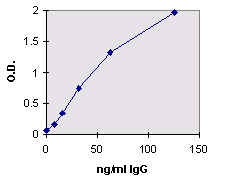
Using linear graph paper or a computer program, plot the optical
densities of each standard on the Y-axis versus the
corresponding concentration of the standards on the X-axis.
The
concentration of human IgG in each diluted specimen may then be
determined manually using a ruler to extrapolate, by linear
regression using a computer program or pocket calculator with a
linear regression function, or by point to point calculation
again using a computer or calculator.
Correct the diluted specimen values by the dilution factor used
to obtain the final concentration of human IgG in the original
specimen.
Typical Standard Curve
Below is an example of a typical standard curve. Variations will
occur laboratory to laboratory due to pipetting, incubator
temperatures, plate readers, etc.
|
Human IgG Standard Concentration |
Optical Density
at 450 nm |
|
125 ng/ml |
1.970 |
|
62.5 ng/ml |
1.320 |
|
31.25 ng/ml |
0.737 |
|
15.6 ng/ml |
0.348 |
|
7.8 ng/ml |
0.154 |
|
0 ng/ml |
0.058 |
|
Substrate Blank |
0.050 |
Caution: This kit is for Research Use Only. It is not to be used
as an in vitro Diagnostic |
|
|
|
|
|
PROCEDURAL
FLOW CHART |
|
PREPARE REAGENT DILUTIONS
PIPET SPECIMENS AND STANDARDS
INCUBATE 30 MINUTES AT 370+ 10C
WASH PLATE
PIPET DETECTOR ANTIBODY
INCUBATE 30 MINUTES AT 370+ 10C
WASH PLATE
PIPET SUBSTRATE SOLUTION
INCUBATE 30 MINUTES AT ROOM TEMPERATURE
ADD STOP SOLUTION AND READ AT 450 NM |
|
|
|
|
Rev. 09/00
|
