Home ] Up ] Immunology Infectious agents Cellular Biology Molecular biology Instruments Chimical Product PCR ELISA Sites Komabiotech products Product NEW antibodies NEW Cells Culturs EPIGENTEK Electrophoresis Contact Us EnoGene SACACE Albumin—OsrHSA GENTAUR NEWS Antibodies & Supporting ToolsHome ] Up ] NAD/NADH Electrophoresis  Albumin—OsrHSA Pricelist:  http://www.diagrade.com/search.php  

Google

       Pricelist 2010 

Home
Up

OUR SUPPLIERS

KOMABIOTECH
301, Gayang Technotown, #1487 Gayang 3 dong, Gangseo-gu
Seoul 157-793, KOREA

Spherotech, Inc.
27845 Irma Lee Circle, Unit 101
Lake Forest, IL 60045

Exalpha Biologicals, Inc.
2 Shaker Road, Unit B101
Shirley, MA 01464

SCETI K.K
BIOSCIENCE
Export                                   DF Kasumigaseki Place,3-6-7 Kasumigaseki Chiyoda-ku, Tokyo 100-0013 JAPAN 

EY Laboratories, Inc. Headquarters
107 N. Amphlett Blvd
San Mateo, CA. 94401 USA

EXBIO Praha, a.s.
Nad Safinou II 366
252 42  Vestec
Czech Republic


Sacace Biotechnologies S.r.l.

Via Scalabrini, 44
22100 Como Italy


redcoon België GENTAUR BVBA

VAT BE0473327336

Av. de l Armee 68 B4

1040 Brussels BELGIUM

  Tel + 32 16 58 90 45 

Fax + 32 16 50 90 45

GENTAUR France SARL

SIRET 48423788800017

Rue Lagrange, 9

75005 Paris, France

 Tel 01 43 25 01 50

Fax 01 43 25 01 60 

GENTAUR Germany Marienbongard 20

52074 Aachen, Germany

Tel  0241 56 00 99 68                    Fax 0241 56 00 47 88 

GENTAUR Pol Sp. Z.o.o. Ulica Ogarna 15/19B m2

80-826 GDANSK

Tel 00 48 58 760 77 08

Fax: 00 32 16 50 90 45

GENTAUR Italy

23015 Milano, Italy

 Tel 02 36 00 65 93

Fax 02 36 00 65 94

Česká republika Praha
+420246019719
 

Danmark

+4569918806 

Finland Helsset
+358942419041

Ελλάς Αθήνα
+302111768494
 

Ireland Dublin
+35316526556
 

Luxembourg
+35220880274
 

Magyarország Budapest
+3619980547
 

Nederland
+31208080893
 

Norge Oslo
+4721031366
 

Österreich
+43720880899
 

Sverige Stockholm
+46852503438
 

Schweiz Züri
+41435006251


Northern America 

Canada Montreal
+15149077481
 

US New York
+17185132983


Other Countries redcoon België
0032 (0)16 41 44 07


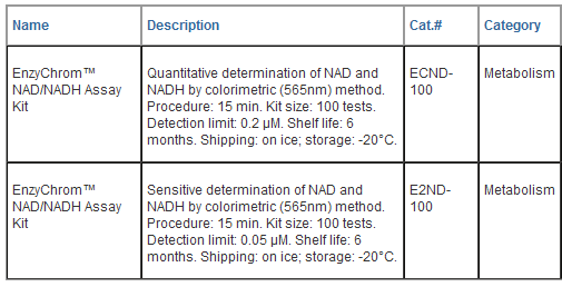
EnzyChromTM NAD+/NADH Assay Kit (ECND-100)

Ultrasensitive Colorimetric Determination of NAD+/NADH at 565 nm

DESCRIPTION

Pyridine nucleotides play an important role in metabolism and, thus, there is continual interest in monitoring their concentration levels. Quantitative determination of NAD+/NADH has applications in research pertaining to energy transformation and redox state of cells or tissue. Simple, direct and automation-ready procedures for measuring NAD+/NADH concentration are very desirable. BioAssay Systems' EnzyChromTM NAD+/NADH assay kit is based on an alcohol dehydrogenase cycling reaction, in which a tetrazolium dye (MTT) is reduced by NADH in the presence of phenazine methosulfate (PMS). The intensity of the reduced product color, measured at 565 nm, is proportionate to the NAD+/NADH concentration in the sample. This assay is highly specific for NAD+/NADH and is not interfered by NADP+/NADPH. Our assay is a convenient method to measure NAD, NADH and their ratio.

APPLICATIONS

Direct Assays: NAD+/NADH concentrations and ratios in cell or tissue extracts.

KEY FEATURES

Sensitive and accurate. Detection limit 0.2 μM, linearity up to 10 μM NAD+/NADH in 96-well plate assay.

Convenient. The procedure involves adding a single working reagent, and reading the optical density at time zero and 15 min at room temperature. No 37°C heater is required.

High-throughput. Can be readily automated as a high-throughput 96- well plate assay for thousands of samples per day.

Storage conditions. Store all reagents at -20°C. Shelf life of at least 6 months (see expiry dates on labels).

Precautions: reagents are for research use only. Normal precautions for laboratory reagents should be exercised while using the reagents. Please refer to Material Safety Data Sheet for detailed information.

PROCEDURES

1. Sample Preparation. For tissues weigh ~20 mg tissue for each sample, wash with cold PBS. For cell samples, wash cells with cold PBS and pellet ~105 cells for each sample. Homogenize samples (either tissue or cells) in a 1.5 mL eppindorf tube with either 100 μL NAD extraction buffer for NAD determination or 100 μL NADH extraction buffer for NADH determination. Heat extracts at 60°C for 5 min and then add 20 μL Assay Buffer and 100 μL of the opposite extraction buffer to neutralize the extracts. Briefly vortex and spin the samples down at 14,000 rpm for 5 min. Use supernatant for NAD/NADH assays. Determination of both NAD and NADH concentrations requires extractions from two separate samples.

2. Calibration Curve. Prepare 500 μL 10 μM NAD Premix by mixing 5 μL 1 mM Standard and 495 μL distilled water. Dilute standard as follows.

Transfer 40 μL standards into wells of a clear bottom 96-well plate.

Samples. Add 40 μL sample per well in separate wells.

3. Reagent Preparation. For each well of reaction, prepare Working Reagent by mixing 50 μL Assay Buffer, 1 μL Enzyme, 10 μL 1v% Ethanol, 14 μL PMS and 14 μL MTT. Fresh reconstitution is recommended.

4. Reaction. Add 80 μL Working Reagent per well quickly. Tap plate to mix briefly and thoroughly.

5. Read optical density (OD0) for time “zero” at 565 nm (520-600nm) and OD15 after a 15-min incubation at room temperature.

6. Calculation. Subtract OD0 from OD15 for the standard and sample wells. Use the DOD values to determine sample NAD/NADH concentration from the standard curve.

Note: If the sample DOD values are higher than the DOD value for the 10 μM standard, dilute sample in distilled water and repeat this assay. Multiply the results by the dilution factor.

MATERIALS REQUIRED, BUT NOT PROVIDED

Pipetting (multi-channel) devices. Clear-bottom 96-well plates (e.g. Corning Costar) and plate reader.

GENERAL CONSIDERATIONS

1. At these concentrations, the standard curves for NAD and NADH are identical. Since NADH in solution is unstable, we provide only NAD as the standard.

2. This assay is based on an enzyme-catalyzed kinetic reaction. Addition of Working Reagent should be quick and mixing should be brief but thorough. Use of multi-channel pipettor is recommended.

3. The following substances interfere and should be avoided in sample preparation. Ascorbic acid, SDS (>0.2%), sodium azide, NP-40 (>1%) and Tween-20 (>1%).

LITERATURE

1. Zhao, Z, Hu, X and Ross CW (1987). Comparison of Tissue Preparation Methods for Assay of Nicotinamide Coenzymes. Plant Physiol. 84: 987-988.

2. Matsumura, H. and Miyachi S (1980). Cycling assay for nicotinamide adenine dinucleotides. Methods Enzymol. 69: 465-470.

3. Vilcheze, C et al. (2005). Altered NADH/NAD+ Ratio Mediates Coresistance to Isoniazid and Ethionamide in Mycobacteria. Antimicrobial Agents and Chemotherapy. 49(2): 708-720.


EnzyChromTM NAD+/NADH Assay Kit (E2ND-100)

Ultrasensitive Colorimetric Determination of NAD+/NADH at 565 nm

DESCRIPTION

Pyridine nucleotides play an important role in metabolism and, thus, there is continual interest in monitoring their concentration levels. Quantitative determination of NAD+/NADH has applications in research pertaining to energy transformation and redox state of cells or tissue. Simple, direct and automation-ready procedures for measuring NAD+/NADH concentration are very desirable. BioAssay Systems' EnzyChromTM NAD+/NADH assay kit is based on a lactate dehydrogenase cycling reaction, in which the formed NADH reduces a formazan (MTT) reagent. The intensity of the reduced product color, measured at 565 nm, is proportionate to the NAD+/NADH concentration in the sample. This assay is highly specific for NAD+/NADH and with minimal interference (<1%) by NADP+/NADPH. Our assay is a convenient method to measure NAD, NADH and their ratio.

APPLICATIONS

Direct Assays: NAD+/NADH concentrations and ratios in cell or tissue extracts.

KEY FEATURES

Sensitive and accurate. Detection limit 0.05 μM, linearity up to 10 μM NAD+/NADH in 96-well plate assay.

Convenient. The procedure involves adding a single working reagent, and reading the optical density at time zero and 15 min at room temperature. No 37°C heater is required.

High-throughput. Can be readily automated as a high-throughput 96- well plate assay for thousands of samples per day.

Storage conditions. Store all reagents at -20°C. Shelf life of at least 6 months after receipt.

Precautions: reagents are for research use only. Normal precautions for laboratory reagents should be exercised while using the reagents. Please refer to Material Safety Data Sheet for detailed information.

PROCEDURES

1. Sample Preparation. For tissues weigh ~20 mg tissue for each sample, wash with cold PBS. For cell samples, wash cells with cold PBS and pellet ~105 cells for each sample. Homogenize samples (either tissue or cells) in a 1.5 mL Eppindorf tube with either 100 μL NAD extraction buffer for NAD determination or 100 μL NADH extraction buffer for NADH determination. Heat extracts at 60°C for 5 min and then add 20 μL Assay Buffer and 100 μL of the opposite extraction buffer to neutralize the extracts. Briefly vortex and spin the samples down at 14,000 rpm for 5 min. Use supernatant for NAD/NADH assays. Determination of both NAD and NADH concentrations requires extractions from two separate samples.

2. Calibration Curve. Prepare 500 μL 10 μM NAD Premix by mixing 5 μL 1 mM Standard and 495 μL distilled water. Dilute standard as follows.

Transfer 40 μL standards into wells of a clear flat-bottom 96-well plate.

Samples. Add 40 μL sample per well in separate wells.

3. Reagent Preparation. For each well of reaction, prepare Working Reagent by mixing 60 μL Assay Buffer, 1 μL Enzyme A, 1 μL Enzyme B, 14 μL Lactate and 14 μL MTT. Fresh reconstitution is recommended.

4. Reaction. Add 80 μL Working Reagent per well quickly. Tap plate to mix briefly and thoroughly.

5. Read optical density (OD0) for time “zero” at 565 nm (520-600nm) and OD15 after a 15-min incubation at room temperature.

6. Calculation. Subtract OD0 from OD15 for the standard and sample wells. Use the DOD values to determine sample NAD/NADH concentration from the standard curve.

Note: If the sample DOD values are higher than the DOD value for the 10 μM standard, dilute sample in distilled water and repeat this assay. Multiply the results by the dilution factor.

MATERIALS REQUIRED, BUT NOT PROVIDED

Pipetting (multi-channel) devices. Clear-bottom 96-well plates (e.g. Corning Costar) and plate reader.

GENERAL CONSIDERATIONS

1. At these concentrations, the standard curves for NAD and NADH are identical. Since NADH in solution is unstable, we provide only NAD as the standard.

2. This assay is based on an enzyme-catalyzed kinetic reaction. Addition of Working Reagent should be quick and mixing should be brief but thorough. Use of multi-channel pipettor is recommended.

3. The following substances interfere and should be avoided in sample preparation. EDTA (>0.5 mM), ascorbic acid, SDS (>0.2%), sodium azide, NP-40 (>1%) and Tween-20 (>1%).

LITERATURE

1. Zhao, Z, Hu, X and Ross CW (1987). Comparison of Tissue Preparation Methods for Assay of Nicotinamide Coenzymes. Plant Physiol. 84: 987-988.

2. Matsumura, H. and Miyachi S (1980). Cycling assay for nicotinamide adenine dinucleotides. Methods Enzymol. 69: 465-470.

3. Vilcheze, C et al. (2005). Altered NADH/NAD+ Ratio Mediates Coresistance to Isoniazid and Ethionamide in Mycobacteria. Antimicrobial Agents and Chemotherapy. 49(2): 708-720.