|
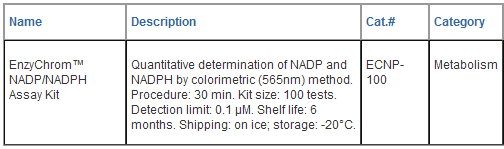
EnzyChromTM NADP+/NADPH Assay Kit
(ECNP-100)
Ultrasensitive Colorimetric
Determination of NADP+/NADPH at 565 nm
DESCRIPTION
Pyridine nucleotides play an
important role in metabolism and, thus, there is
continual interest in monitoring their concentration
levels. Quantitative determination of NADP+/NADPH has
applications in research pertaining to energy
transformation and redox state of cells or tissue.
Simple, direct and automation-ready procedures for
measuring NADP+/NADPH concentration are very desirable.
BioAssay Systems' EnzyChromTM NADP+/NADPH assay kit is
based on a glucose dehydrogenase cycling reaction, in
which the formed NADPH reduces a formazan (MTT) reagent.
The intensity of the reduced product color, measured at
565 nm, is proportionate to the NADP+/NADPH
concentration in the sample. This assay is highly
specific for NADP+/NADPH and is not interfered by
NAD+/NADH. Our assay is a convenient method to measure
NADP, NADPH and their ratio.
APPLICATIONS
Direct
Assays:
NADP+/NADPH concentrations and ratios in cell or tissue
extracts.
KEY
FEATURES
Sensitive
and accurate.
Detection limit 0.1 μM, linearity up to 10 μM
NADP+/NADPH in 96-well plate assay.
Convenient. The procedure involves adding a single
working reagent, and reading the optical density at time
zero and 30 min at room temperature. No 37°C heater is
required.
High-throughput. Can be readily automated as a
high-throughput 96- well plate assay for thousands of
samples per day.
KIT
CONTENTS (100 tests in 96-well plates)
Assay
Buffer: 10 mL Glucose (1 M): 1.5 mL
MTT
Solution: 1.5 mL Enzyme Mix: 120
μL
NADP
Standard: 0.5 mL 1 mM
NADP/NADPH
Extraction Buffers: each 12 mL
Storage
conditions.
Store all reagents at -20°C. Shelf life of at least 6
months (see expiry dates on labels).
Precautions: reagents are for research use only.
Normal precautions for laboratory reagents should be
exercised while using the reagents. Please refer to
Material Safety Data Sheet for detailed information.
PROCEDURES
1.
Sample Preparation. For tissues weigh ~20 mg tissue
for each sample, wash with cold PBS. For cell samples,
wash cells with cold PBS and pellet ~105 cells for each
sample. Homogenize samples (either tissue or cells) in a
1.5 mL eppindorf tube with either 100 μL NADP extraction
buffer for NADP determination or 100 μL NADPH extraction
buffer for NADPH determination. Heat extracts at 60°C
for 5 min and then add 20 μL Assay Buffer and 100 μL of
the opposite extraction buffer to neutralize the
extracts. Briefly vortex and spin the samples down at
14,000 rpm for 5 min. Use supernatant for NADP/NADPH
assays. Determination of both NADP and NADPH
concentrations requires extractions from two separate
samples.
2.
Calibration Curve. Prepare 500 μL 10 μM NADP Premix
by mixing 5 μL 1 mM Standard and 495 μL distilled water.

Dilute standard as shown in the
Table. Transfer 40 μL standards into wells of a clear
bottom 96-well plate. Samples: add 40 μL sample
per well in separate wells. 3. Reagent Preparation.
For best results allow Enzyme to come to RT (15-30 min)
before preparing the Working Reagent. For each well of
reaction, prepare Working Reagent by mixing 60 μL Assay
Buffer, 1 μL Enzyme Mix, 10 μL Glucose and 14 μL MTT.
Fresh reconstitution is recommended.
4. Reaction. Add 80 μL Working
Reagent per well quickly. Tap plate to mix briefly and
thoroughly.
5. Read optical density (OD0) for
time “zero” at 565 nm (520-600nm) and OD30 after a
30-min incubation at room temperature.
6. Calculation. Subtract OD0
from OD30 for the standard and sample wells. Use the DOD
values to determine sample NADP/NADPH concentration from
the standard curve. Note: If the sample DOD values are
higher than the DOD value for the 10 μM standard, dilute
sample in distilled water and repeat this assay.
Multiply the results by the dilution factor.
MATERIALS REQUIRED, BUT NOT PROVIDED
Pipetting (multi-channel) devices.
Clear-bottom 96-well plates (e.g. Corning Costar) and
plate reader.
GENERAL CONSIDERATIONS
1. At these concentrations, the
standard curves for NADP and NADPH are identical. Since
NADPH in solution is unstable, we provide only NADP as
the standard.
2. This assay is based on an
enzyme-catalyzed kinetic reaction. Addition of Working
Reagent should be quick and mixing should be brief but
thorough. Use of multi-channel pipettor is recommended.
3. The following substances interfere
and should be avoided in sample preparation. EDTA (>0.5
mM), ascorbic acid, SDS (>0.2%), sodium azide, NP-40
(>1%) and Tween-20 (>1%).

PUBLICATIONS
1. Ding X et al (2009). Enhanced
HtrA2/Omi expression in oxidative injury to retinal
pigment epithelial cells and murine models of
neurodegeneration. Invest Ophthalmol Vis Sci.
50(10):4957-66.
2. Tseng HC et al (2009). Metabolic
engineering of Escherichia coli for enhanced production
of (R)- and (S)-3-hydroxybutyrate. Appl Environ
Microbiol. 75(10):3137-45.
3. Du J et al (2010). Mechanisms of
ascorbate-induced cytotoxicity in pancreatic cancer.
Clin Cancer Res. 16(2):509-20.
|





















 GENTAUR Germany
Marienbongard 20
GENTAUR Germany
Marienbongard 20
 GENTAUR Italy
 Česká republika
Praha
Česká republika
Praha Danmark
 Finland Helsset
 Ελλάς Αθήνα
 Ireland Dublin
 Luxembourg
 Magyarország
Budapest
Magyarország
Budapest Nederland
 Norge
Oslo
Norge
Oslo Österreich
 Sverige
Stockholm
Sverige
Stockholm Schweiz Züri
 Canada Montreal
 US New York
 日本
日本