|


|
OUR SUPPLIERS
KOMABIOTECH
301,
Gayang Technotown, #1487
Gayang 3 dong,
Gangseo-gu
Seoul 157-793, KOREA
Spherotech, Inc.
27845 Irma Lee Circle, Unit 101
Lake Forest, IL 60045
|
|
Exalpha
Biologicals,
Inc.
2 Shaker Road,
Unit B101
Shirley, MA
01464
SCETI K.K
BIOSCIENCE Export DF Kasumigaseki Place,3-6-7 Kasumigaseki Chiyoda-ku, Tokyo 100-0013 JAPAN
|
EY
Laboratories, Inc. Headquarters
107 N.
Amphlett Blvd
San Mateo, CA. 94401 USA
EXBIO Praha, a.s.
Nad
Safinou II 366
252 42 Vestec
Czech Republic
Sacace Biotechnologies S.r.l.
Via Scalabrini,
44
22100 Como Italy
 GENTAUR BVBA
VAT BE0473327336
Av. de l Armee 68 B4
1040 Brussels
BELGIUM
Tel + 32 16 58 90
45
Fax + 32 16 50 90 45
 GENTAUR France SARL
SIRET 48423788800017
Rue Lagrange, 9
75005 Paris,
France
Tel 01 43 25 01 50
Fax 01 43 25 01 60
 GENTAUR Germany
Marienbongard 20
GENTAUR Germany
Marienbongard 20
52074 Aachen,
Germany
Tel 0241 56 00
99 68
Fax 0241 56 00 47 88
 GENTAUR
Pol Sp. Z.o.o. Ulica
Ogarna 15/19B m2 GENTAUR
Pol Sp. Z.o.o. Ulica
Ogarna 15/19B m2
80-826 GDANSK
Tel 00 48 58 760 77
08
Fax: 00 32 16 50 90
45
 GENTAUR Italy
23015 Milano, Italy
Tel 02 36 00 65
93
Fax 02 36 00 65
94
 Česká republika
Praha
Česká republika
Praha
+420246019719
 Danmark
+4569918806
 Finland Helsset
+358942419041
 Ελλάς Αθήνα
+302111768494
 Ireland Dublin
+35316526556
 Luxembourg
+35220880274
 Magyarország
Budapest
Magyarország
Budapest
+3619980547
 Nederland
+31208080893
 Norge
Oslo Norge
Oslo
+4721031366
 Österreich
+43720880899
 Sverige
Stockholm Sverige
Stockholm
+46852503438
 Schweiz Züri
+41435006251
Northern America
 Canada Montreal
+15149077481
 US New York
+17185132983
Other Countries
0032 (0)16 41 44 07 |
|
|
|
|
|
|

| |
|
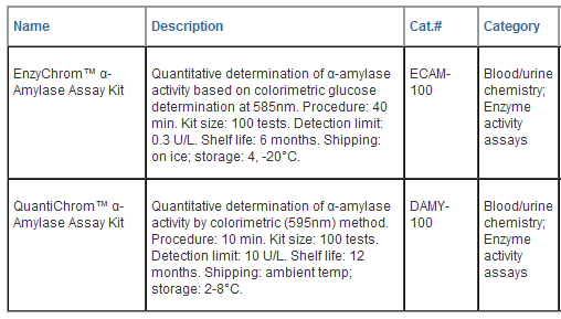
EnzyChromTM
a-Amylase Assay Kit (ECAM-100)
Quantitative Colorimetric Amylase Determination at
585nm
DESCRIPTION
AMYLASE belongs to the family of glycoside
hydrolase enzymes that break down starch into glucose molecules by
acting on α-1,4-glycosidic bonds. The α-amylases (EC 3.2.1.1) cleave at
random locations on the starch chain, ultimately yielding maltotriose
and maltose, glucose and "limit dextrin" from amylose and amylopectin.
In mammals, α-amylase is a major digestive enzyme. Increased enzyme
levels in humans are associated with salivary trauma, mumps due to
inflammation of the salivary glands, pancreatitis and renal failure.
Simple, direct and automation-ready procedures for measuring amylase
activity are very desirable. BioAssay Systems’ EnzyChromTM a-amylase
assay method involves two steps: (1). α-amylase in the sample hydrolyzes
starch and the product is rapidly converted to glucose by α-glucosidase
and hydrogen peroxide by glucose oxidase; (2). hydrogen peroxide
concentration is determined with a colorimetric reagent.
APPLICATIONS
Direct assays of
α-amylase activity in
blood, saliva, urine, grains and
other agricultural
samples.
KEY FEATURES
Sensitive and accurate .
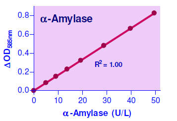
Linear detection range 0.3 to 50 U/L α- amylase in 96-well plate assay.
Convenient. The procedure involves adding a single working reagent,
incubation for 15 min, followed by the detection reagent and a 20-min
incubation and reading the optical density at 585 nm.
KIT CONTENTS
Assay Buffer (pH 7.0): 20 mL Substrate: 120
μL
Detection Reagent: 20 mL Enzyme A: 120
μL
Glucose Standard: 1 mL Enzyme B: 120
μL
Storage conditions .
Kit is shipped on ice. Store Detection Reagent at
4°C
and others at -20°C. Shelf life of at least 6 months (see expiry
dates on labels).
Precautions: reagents are for research use only.
Normal precautions for laboratory reagents should be exercised while
using the reagents. Please refer to Material Safety Data Sheet for
detailed information.
PROCEDURES
Reagents. Equilibrate all components to room
temperature. Keep thawed Enzyme Mix in a refrigerator or on ice. The
substrate may have precipitates. Prior to use, vortex tube to dissolve
precipitates; gentle swirl the Detection Reagent bottle .
Sample preparation.
Ideally samples are assayed fresh. When stored
frozen,
α-amylase is stable for one
month. Ascorbic acid, heparin,
EDTA, EGTA, citrate, SDS,
Tris (> 8mM) and ethanol (>0.4%) interfere and should be avoided in
sample preparation. If glucose is present in the sample, treat the
samples as described in GENERAL CONSIDERATIONS. It is prudent to perform
a pilot test with samples at various dilutions. Recommended dilution:
serum 50-fold, saliva 2,000-fold in Assay Buffer prior to assay.
1.
Prepare 400 μM
Glucose Standard by mixing 10 μL of the provided
(300 mg/dL) standard with
406 μL Assay Buffer.
Transfer 10 μL
Assay Buffer, 10 μL
400 μM glucose, and 10 μL of each sample
into separate wells of a
clear flat-bottom 96-well plate.
2. Prepare an appropriate Working Reagent for each well.
For Standards and samples: mix 40
μL Assay Buffer, 0.5 μL
Substrate, 1
μL Enzyme A, 1 μL
Enzyme B. Transfer
40 μL working
reagent and blank control reagent to the
appropriate sample and
blank wells. Tap plate to mix. Incubate for 15 min at room temperature
(25°C).
3. Add 150
μL Detection Reagent to
each well. Mix and incubate for 20
min at room temperature
(25°C). Read OD585nm
(540-610nm) on a
plate reader.
CALCULATION
The Amylase activity is calculated as

OD SAMPLE,
ODSTD and ODBUFFER are optical density values of the
sample, the 400
μM glucose standard
and Assay Buffer. t is the
incubation time.
t = 15 min in the
standard protocol.
n is
the dilution
factor (n =
50 for serum, 2000 for saliva). One unit of enzyme
catalyzes the production
of 1 μmole of
glucose per min under the
assay conditions.
Note: if the calculated activity is higher than 50 U/L,
dilute sample in Assay Buffer and repeat assay. Multiply the results by
the dilution factor.
MATERIALS REQUIRED, BUT NOT PROVIDED
Pipeting devices, centrifuge tubes, clear flat-bottom
96-well plates, plate reader, and optionally membrane filters (e.g.
Microcon YM-10 from Millipore).
GENERAL CONSIDERARIONS
For samples known to contain glucose, use a membrane
filter (e.g. Microcon YM-10 from Millipore) to remove glucose: load 50
μL sample in
a Microcon YM-10 (10 kDa
cutoff) and add 500
μL Assay Buffer.
Centrifuge at 14000 rpm for 30 min, check level of sample, ideally the
sample level will be less than 50
μL. Add 500 μL Assay Buffer
and repeat the
centrifugation. Measure final sample volume with a pipetman and
calculate dilution factor
n = final sample
volume/50 μL.
LITERATURE
1. Gullbault, GG and Rietz, E.B. (1976). Enzymatic,
Fluorometric Assay of a-Amylase in Serum. Clin. Chem. 22(10): 1702:1704.
2. Mashige F, Ohkubo A, Kamei S, Yamanaka M. (1982). An
enzymic method for urine and serum alpha-amylase assay. Clin. Chem.
28(7):1715-6.
3. Marshall JJ, Iodice AP, Whelan WJ. (1977). A new
serum alphaamylase assay of high sensitivity. Clin Chim Acta.
76(2):277-83.
|
QuantiChromTM a-Amylase Assay Kit (DAMY-100)
Rapid Colorimetric Determination of
α-Amylase
Activity at 595nm
DESCRIPTION
AMYLASE belongs to the family of
glycoside hydrolase enzymes that break down starch into glucose
molecules by acting on
α-1,4-glycosidic
bonds. The
α-amylases
(EC 3.2.1.1) cleave at random locations on the starch chain,
ultimately yielding maltotriose and maltose, glucose and "limit
dextrin" from amylose and amylopectin. In mammals,
α-amylase
is a major digestive enzyme. Increased enzyme levels in humans
are associated with salivary trauma, mumps due to inflammation
of the salivary glands, pancreatitis and renal failure. Simple,
direct and automation-ready procedures for measuring
α-amylase
activity are very desirable. BioAssay Systems' QuantiChromTM
α-amylase
Assay uses an insoluble dye-coupled substrate amylose azure,
which is cleaved by
α-amylase
into soluble colored products. The color intensity, measured at
595 nm, is proportionate to the enzyme activity in the sample.
APPLICATIONS
Direct assays of
α-amylase
activity in serum, heparinized plasma, saliva, urine and other
biological samples.
KEY FEATURES
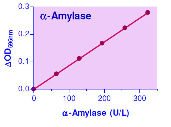
Sensitive and accurate. Linear detection
range 2 to 300 U/L
α-amylase
in 96-well plate assay.
Convenient. The procedure involves adding
a single working reagent, and reading the optical density at 5
min at room temperature or 37°C.
KIT CONTENTS
Substrate (pH 7.0): 20 mL
Stop Reagent: 10 mL
Calibrator: 2 mL (equivalent to 550 U/L)
Storage conditions. Store all reagents at
2-8°C. Shelf life of at least 6 months (see expiry dates on
labels).
Precautions: reagents are for research
use only. Normal precautions for laboratory reagents should be
exercised while using the reagents. Please refer to Material
Safety Data Sheet for detailed information.
PROCEDURES
Sample preparation. Ideally samples are
assayed fresh. When stored frozen,
α-amylase
is stable for one month. EDTA, EGTA and citrate are
α-amylase
inhibitors and should be avoided in sample preparation.
1. Reaction. In appropriately labeled
1.5-mL Eppendorf tubes, transfer 10 μL sample. Add 190 μL
Substrate, vortex briefly to mix and incubate for 5 min.
Caution: the substrate contains blue
insoluble materials that tend to settle. It is important to
invert the bottle ten times to maintain the substrate in
suspension and then quickly transfer the substrate into sample
tubes. If the assay is to be performed at 37°C, warm up
the Substrate to this temperature prior to sample addition.
As a blank control, use 10 μL water plus 190
μL Substrate and 80 μL Stop Reagent. Alternatively a blank
control can be prepared in the order: 10 μL sample, 80 μL Stop
Reagent and 190 μL Substrate. This would be necessary if the
sample has a background color that is visibly blue.
2. Add 80 μL Stop Reagent to each sample tube
to terminate the reaction. Vortex to mix and centrifuge for 5
min at 14,000 rpm. Carefully transfer 200 μL supernatant into
wells of a clear bottom 96-well plate. In separate wells,
transfer 200 μL water and 200 μL Calibrator.
3. Read OD595nm (580 to 600nm) on a plate
reader.
4. Calculation:
α -amylase
activity is calculated as follows

where ODSAMPLE and ODBLANK are the OD595nm
values of the sample and blank, respectively. ODCAL and ODH2O
are the OD595nm values of the Calibrator and water. n is
the dilution factor. The number “550” is the equivalent activity
(U/L) of the calibrator under the assay conditions.
Note: if the calculated activity is
higher than 300 U/L, dilute sample in water and repeat assay.
Multiply the results by the dilution factor (n).
Unit definition: one unit of enzyme
catalyzes the production of 1 μmole of product per minute under
the assay conditions (pH 7.0).
MATERIALS REQUIRED, BUT NOT PROVIDED
Pipeting (multi-channel) devices. 1.5-mL
Eppendorf tubes. Centrifuge. Clear-bottom 96-well plates (e.g.
Corning Costar) and plate reader.
GENERAL CONSIDERATIONS
For assays in standard 1 mL cuvet, use 1 mL
water and 1 mL Calibrator. Sample reactions: 40 μL sample + 720
μL Substrate and 320 μL Stop Reagent, after incubation and
centrifugation, remove 1 mL for OD determination.
EXAMPLES
Samples were assayed in duplicate using the
96-well plate protocol.
α-amylase
activities were 149 ± 3 U/L for rat plasma, 172 ± 6 U/L for rat
serum, 164 ± 2 U/L for mouse serum, 28.4 ± 0.8 U/L for human
serum, 29.9 ± 0.4 U/L for human plasma, 27.9 ± 0.4 U/L for human
urine and 10,578 ± 390 U/L for a human saliva sample.
LITERATURE
1. Rinderknecht H, Wilding P, Haverback BJ.
(1967) A new method for the determination of alpha-amylase.
Experientia. 23(10):805
2. Klein B, Foreman JA. (1980) Amylolysis of a
chromogenic substrate, Cibachron Blue F3GA-amylose: kinetics and
mechanism. Clin Chem. 26(2):250-3.
3. Klein B, Foreman JA, Searcy RL. (1970) New
chromogenic substrate for determination of serum amylase
activity. Clin Chem. 16(1):32-8. |
|
|




















 日本
日本