|


|
OUR SUPPLIERS
KOMABIOTECH
301,
Gayang Technotown, #1487
Gayang 3 dong,
Gangseo-gu
Seoul 157-793, KOREA
Spherotech, Inc.
27845 Irma Lee Circle, Unit 101
Lake Forest, IL 60045
|
|
Exalpha
Biologicals,
Inc.
2 Shaker Road,
Unit B101
Shirley, MA
01464
SCETI K.K
BIOSCIENCE Export DF Kasumigaseki Place,3-6-7 Kasumigaseki Chiyoda-ku, Tokyo 100-0013 JAPAN
|
EY
Laboratories, Inc. Headquarters
107 N.
Amphlett Blvd
San Mateo, CA. 94401 USA
EXBIO Praha, a.s.
Nad
Safinou II 366
252 42 Vestec
Czech Republic
Sacace Biotechnologies S.r.l.
Via Scalabrini,
44
22100 Como Italy
 GENTAUR BVBA
VAT BE0473327336
Av. de l Armee 68 B4
1040 Brussels
BELGIUM
Tel + 32 16 58 90
45
Fax + 32 16 50 90 45
 GENTAUR France SARL
SIRET 48423788800017
Rue Lagrange, 9
75005 Paris,
France
Tel 01 43 25 01 50
Fax 01 43 25 01 60
 GENTAUR Germany
Marienbongard 20
GENTAUR Germany
Marienbongard 20
52074 Aachen,
Germany
Tel 0241 56 00
99 68
Fax 0241 56 00 47 88
 GENTAUR
Pol Sp. Z.o.o. Ulica
Ogarna 15/19B m2 GENTAUR
Pol Sp. Z.o.o. Ulica
Ogarna 15/19B m2
80-826 GDANSK
Tel 00 48 58 760 77
08
Fax: 00 32 16 50 90
45
 GENTAUR Italy
23015 Milano, Italy
Tel 02 36 00 65
93
Fax 02 36 00 65
94
 Česká republika
Praha
Česká republika
Praha
+420246019719
 Danmark
+4569918806
 Finland Helsset
+358942419041
 Ελλάς Αθήνα
+302111768494
 Ireland Dublin
+35316526556
 Luxembourg
+35220880274
 Magyarország
Budapest
Magyarország
Budapest
+3619980547
 Nederland
+31208080893
 Norge
Oslo Norge
Oslo
+4721031366
 Österreich
+43720880899
 Sverige
Stockholm Sverige
Stockholm
+46852503438
 Schweiz Züri
+41435006251
Northern America
 Canada Montreal
+15149077481
 US New York
+17185132983
Other Countries
0032 (0)16 41 44 07 |
|
|
|
|
|
|

| |
|
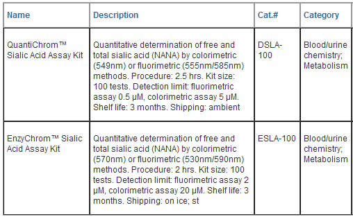
QuantiChromTM Sialic Acid
Assay Kit (Cat# DSLA-100)
Quantitative Determination of
Free and Total Sialic Acid
DESCRIPTION
SIALIC ACID is a general
name for nine carbon acidic sugars with N- or
O-substituted derivatives. The most common
member of these sugars is N-acetylneuraminic
acid (NANA). Sialic acid is widely distributed
throughout mammalian tissues and fluids
including serum. Sialylated oligosaccharides
have been shown to exhibit antiviral properties
and are also known to influence blood
coagulation and cholesterol levels. The sialic
acid level in body fluids is also an important
marker for diagnosing cancer. Simple and direct
procedures for measuring sialic acid
concentrations find wide applications in
research and drug discovery. BioAssay Systems'
sialic acid assay uses an improved Warren
method, in which sialic acid is oxidized to
formylpyruvic acid which reacts with
thiobarbituric acid to form a pink colored
product. The color intensity at 549 nm or
fluorescence intensity at lem/ex = 585/555 nm is
directly proportional to sialic acid
concentration in the sample.
KEY FEATURES
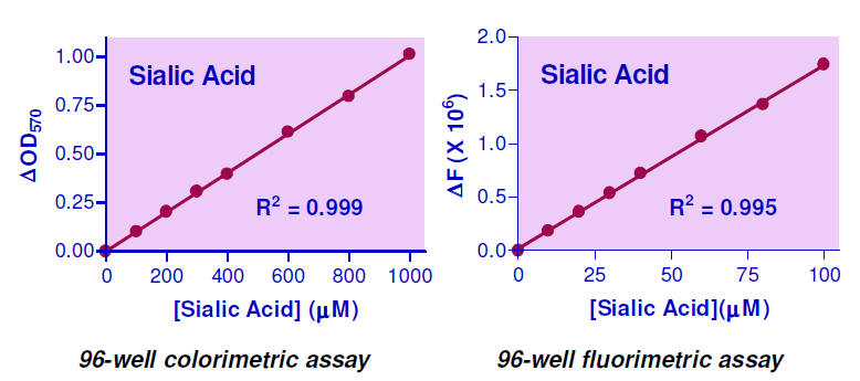
Sensitive and accurate .
Use as little as 60 μL samples. Linear detection
range in 96-well plate: 5 to 1000 μM sialic acid
for colorimetric assays and 0.5 to 100 μM for
fluorimetric assays.
APPLICATIONS:
Direct Assays:
sialic acid in biological samples (e.g. serum,
plasma, saliva, milk).
KIT CONTENTS
Dye Reagent:
6
mL Oxidation Reagent: 10 mL
10% TCA: 5 mL
Hydrolysis Reagent: 10 mL
DMSO: 12 mL Standard:
500 μL 10 mM Sialic Acid
Storage conditions. The
kit is shipped at ambient temperature. Store the
Standard at -20°C, all others at room
temperature. Shelf life of three months after
receipt.
Precautions: reagents are
for research use only. Normal precautions for
laboratory reagents should be exercised while
using the reagents. Please refer to Material
Safety Data Sheet for detailed information.
COLORIMTRIC PROCEDURE
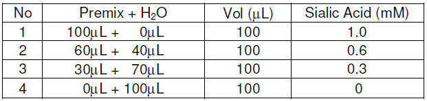
1. Standards.
Equilibrate all components to room temperature.
Prepare a 1000 μM sialic acid standard Premix by
mixing 25 μL of the 10 mM Standard and 225 μL
distilled water dH2O. Dilute Standard as
follows.
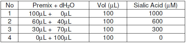
Transfer 20 μL standards into
four labeled Eppendorf tubes, add 5 μL 10% TCA.
2. Samples treatment.
To determine total sialic acid (TSA), samples
need to be hydrolyzed to release bound sialic
acid as follows. In an Eppendorf tube, mix 20 μL
sample, 40 μL dH2O and 40 μL Hydrolysis
Reagent. Heat at 80°C for 60
min, let cool and briefly centrifuge. Add 25 μL
10% TCA, vortex and centrifuge at 14,000 rpm for
10 min. Transfer 25 μL supernatant into a clean
tube and label it “TSA”. To determine free
sialic acid (FSA), directly precipitate protein
by mixing 40 μL sample and 10 μL 10% TCA. Vortex
and centrifuge at 14,000 rpm for 10 min.
Transfer 25 μL supernatant into a clean tube and
label it “FSA”.
3. Oxidation. Prepare
working reagent for each tube by mixing 15 μL
Hydrolysis Reagent, 50 μL dH2O and 65 μL
Oxidation Reagent. Add 125 μL working reagent to
each tube and let stand for 60 min at room
temperature.
4. Color Reaction. Add
50 μL Dye Reagent to each tube. Mix and heat for
10 min at 100°C. Let cool for another 5-10 min.
Add 100 μL DMSO to each tube. Mix and centrifuge
for 5 min at 14,000 rpm. Transfer 250 μL
supernatant into separate wells of a clear,
flat-bottom 96-well plate.
5. Read optical density at
549 nm (540-555nm).
FLUORIMETRIC PROCEDURE
The fluorimetric assay is
10-fold more sensitive than the colorimetric
assay. Prepare standards at 0, 30, 60 and 100 μM
sialic acid in dH2O. The sample treatment,
oxidation and color reaction steps are the same,
except that the final reaction mixture is
transferred into wells of a black, flat-bottom
96-well plate. Read fluorescence intensity at
lex = 555 nm and lem = 585 nm.
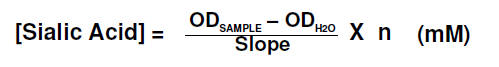
CALCULATION
Subtract blank value (#4)
from the standard values and plot the DOD or DF
against standard concentrations. Determine the
slope and calculate the sialic acid
concentration of Sample,
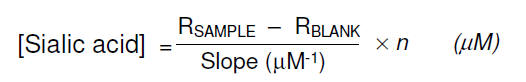
RSAMPLE and RBLANK are
optical density or fluorescence intensity
readings of the Sample and dH2O Blank (#4),
respectively. n is the sample dilution
factor, n = 5 for TSA assays and n
= 1 for FSA assays.
Note: if the Sample OD
value is higher than that for the 1000 μM
Standard, or sample fluorescence intensity
higher than that for the 100 μM Standard, dilute
sample in water and repeat the assay. Multiply
result by the fold of dilution.
Conversions: 1000 μM
NANA equals 30.9 mg/dL or 309 ppm.
MATERIALS REQUIRED, BUT
NOT PROVIDED
Pipeting devices, centrifuge
tubes, centrifuge, heat block, clear flatbottom
96-well plates, black 96-well plates (e.g.
Corning Costar) and plate readers.
LITERATURE
1. Warren, L. (1959). The
Thiobarbituric Acid Assay of Sialic Acids J.
Biol. Chem. 234: 1971-1975.
2. Stefenelli, N. et al
(1985). Serum sialic acid in malignant tumors,
bacterial infections, and chronic liver
diseases. J Cancer Res Clin Oncol. 109(1):55-59.
3. Sherblom, A.P. et al
(1988). Bovine serum sialic acid: age-related
changes in type and content. Int J Biochem.
20:1177-1183. |
|
|
|
|
EnzyChromTM Sialic Acid Assay
Kit (Cat# ESLA-100)
Quantitative Colorimetric/Fluorimetric
Sialic Acid Determination
DESCRIPTION
SIALIC ACID
is a general name for nine carbon acidic sugars
with N- or O-substituted derivatives. The most
common member of these sugars is N-acetylneuraminic
acid (NANA). Sialic acid is widely distributed
throughout mammalian tissues and fluids
including serum. Sialylated
oligosaccharides have been shown to exhibit
antiviral properties and are also known to
influence blood coagulation and cholesterol
levels. The Sialic acid level in body fluids is
also an important marker for diagnosing cancer.
Simple, direct and automation-ready procedures
for measuring sialic acid concentrations find
wide applications in research and drug
discovery. BioAssay Systems' sialic acid assay
uses a single Working Reagent that combines NANA
aldolase, pyruvate oxidase and hydrogen peroxide
determination in one step. The color intensity
of the reaction product at 570nm or fluorescence
intensity at lem/ex
= 585/530nm is directly proportional to sialic
acid concentration in the sample.
KEY FEATURES
Sensitive and accurate. Use
as little as 10 μ L
samples. Linear detection range in 96-well
plate: 0.02 to 1 mM sialic acid for colorimetric
assays and 2 to 100
μM
for fluorimetric assays.
Simple and convenient.
Can detect free sialic acid by addition of a
single working reagent and incubation for 60 min
at room temperature or total sialic acid by
pre-treating samples with a 60 min hydrolysis
step.
APPLICATIONS:
Direct Assays: sialic acid in
biological samples.
KIT CONTENTS
Assay Buffer: 10 mL
Hydrolysis Reagent: 10 mL
Enzyme: 120 μ L
Neutralization
Reagent: 5 mL
Dye Reagent: 120 μ L
Standard: 500 μL
10 mM Sialic Acid
Storage conditions.
The kit is shipped on dry ice. Store all
reagents at - 20°C.
Shelf life of three months after receipt.
Precautions:
reagents are for research use only. Normal
precautions for laboratory reagents should be
exercised while using the reagents. Please refer
to Material Safety Data Sheet for detailed
information.
BOUND SIALIC ACID
HYDROLYSIS PROCEDURE
Note :
For measurement of free sialic acid, this
procedure should be skipped.
1. Combine 20
μL
of sample with 80 μL
Hydrolysis Reagent in a microcentrifuge tube
(screw cap tube is preferable) and incubate at
80°C
for 60 min.
2. Allow sample to cool to
room temperature and briefly centrifuge at 14000
rpm to spin down the hydrolysis mixture.
3. Add 20
μL
Neutralization Reagent to each hydrolysis
reaction, briefly vortex to mix and briefly
centrifuge at 14000 rpm to spin down the
reaction. The samples are now ready for the
sialic acid assay.
COLORIMETRIC PROCEDURE
Note :
SH-group containing reagents (e.g.
mercaptoethanol, DTT) may interfere with this
assay and should be avoided in sample
preparation.
1. Equilibrate all components
to room temperature. Prepare a 1 mM Standard
Premix by mixing 50
μL
of the 10 mM Standard and 450
μL
dH2O. Dilute Standard in distilled water as
follows.
Transfer 10
μL
standards and 10 μL
samples into separate wells of a clear
flat-bottom 96-well plate.
2. For each reaction well,
mix 93
μL
Assay Buffer, 1 μL
Dye Reagent and 1 μL
Enzyme in a clean tube. Transfer 90
μL
Working Reagent into each assay well. Tap plate
to mix. Freeze unused reagents for future use.
3. Incubate 60 min at room
temperature. Read optical density at 570nm
(550-585nm).
Note :
if the Sample OD is higher than the Standard OD
at 1 mM,
dilute sample in water and repeat the assay.
Multiply result by the dilution factor.
CALCULATION
Subtract blank OD (water, #4)
from the standard OD values and plot the OD
against standard concentrations. Determine the
slope using linear regression fitting. The
sialic acid concentration of a Sample is
calculated as
where OD SAMPLE
and ODH2O
are the optical density values of the sample and
water, Slope is the slope of the standard curve
in mM-1 and n
is the dilution factor of the sample (n
= 6 for hydrolyzed samples and
n
= 1 for free Sialic Acid samples).
Conversions: 1 mM NANA
equals 30.9 mg/dL or 309 ppm.
FLUORIMETRIC PROCEDURE
1. For fluorimetric assays,
the linear detection range is 2 to 100
μM
sialic acid. Dilute the Standards prepared in
Colorimetric
Procedure 1:10 in H2O.
Transfer 10
μL
standards and 10 μL
samples into separate wells of a
black
96-well plate.
2. Add 90
μL
Working Reagent (see
Colorimetric Procedure).
Tap plate to mix.
3. Incubate 60 min at room
temperature and read fluorescence at
lex
= 530nm and lem
= 585nm.
If assays in 384-well plate
are desired, use 5 μL
Standards and 45 μL
Working Reagent. The sialic acid concentration
of a Sample is calculated as
where F SAMPLE
and FH2O
are the fluorescence values of the sample and
water, Slope is the slope of the standard curve
in μM-1
and n
is the dilution factor of the sample (n
= 6 for hydrolyzed samples and
n = 1 for free
Sialic Acid samples).
MATERIALS REQUIRED, BUT
NOT PROVIDED
Pipeting devices, centrifuge
tubes, Clear flat-bottom 96-well plates, black
96-well or 384-well plates (e.g. Corning Costar)
and plate reader.

LITERATURE
1. Sugahara, K. et al.
(1980). Enzymatic Assay of Serum Sialic Acid.
Clinica Chimica Acta 108:493-8.
2. Simpson, H. et al. (1993).
Serum sialic acid enzymatic assay based on
microtitre plates: application for measuring
capillary serum sialic acid concentrations. Br J
Biomed Sci. 50:164-7. |
|
|
|
|