Home ] Up ] Immunology Infectious agents Cellular Biology Molecular biology Instruments Chimical Product PCR ELISA Sites Komabiotech products Product NEW antibodies NEW Cells Culturs EPIGENTEK Electrophoresis Contact Us EnoGene SACACE Albumin—OsrHSA GENTAUR NEWS Antibodies & Supporting ToolsHome ] Up ] Uric acid Electrophoresis  Albumin—OsrHSA Pricelist:  http://www.diagrade.com/search.php  

Google

       Pricelist 2010 

Home
Up

OUR SUPPLIERS

KOMABIOTECH
301, Gayang Technotown, #1487 Gayang 3 dong, Gangseo-gu
Seoul 157-793, KOREA

Spherotech, Inc.
27845 Irma Lee Circle, Unit 101
Lake Forest, IL 60045

Exalpha Biologicals, Inc.
2 Shaker Road, Unit B101
Shirley, MA 01464

SCETI K.K
BIOSCIENCE
Export                                   DF Kasumigaseki Place,3-6-7 Kasumigaseki Chiyoda-ku, Tokyo 100-0013 JAPAN 

EY Laboratories, Inc. Headquarters
107 N. Amphlett Blvd
San Mateo, CA. 94401 USA

EXBIO Praha, a.s.
Nad Safinou II 366
252 42  Vestec
Czech Republic


Sacace Biotechnologies S.r.l.

Via Scalabrini, 44
22100 Como Italy


redcoon België GENTAUR BVBA

VAT BE0473327336

Av. de l Armee 68 B4

1040 Brussels BELGIUM

  Tel + 32 16 58 90 45 

Fax + 32 16 50 90 45

GENTAUR France SARL

SIRET 48423788800017

Rue Lagrange, 9

75005 Paris, France

 Tel 01 43 25 01 50

Fax 01 43 25 01 60 

GENTAUR Germany Marienbongard 20

52074 Aachen, Germany

Tel  0241 56 00 99 68                    Fax 0241 56 00 47 88 

GENTAUR Pol Sp. Z.o.o. Ulica Ogarna 15/19B m2

80-826 GDANSK

Tel 00 48 58 760 77 08

Fax: 00 32 16 50 90 45

GENTAUR Italy

23015 Milano, Italy

 Tel 02 36 00 65 93

Fax 02 36 00 65 94

Česká republika Praha
+420246019719
 

Danmark

+4569918806 

Finland Helsset
+358942419041

Ελλάς Αθήνα
+302111768494
 

Ireland Dublin
+35316526556
 

Luxembourg
+35220880274
 

Magyarország Budapest
+3619980547
 

Nederland
+31208080893
 

Norge Oslo
+4721031366
 

Österreich
+43720880899
 

Sverige Stockholm
+46852503438
 

Schweiz Züri
+41435006251


Northern America 

Canada Montreal
+15149077481
 

US New York
+17185132983


Other Countries redcoon België
0032 (0)16 41 44 07


QuantiChromTM TBARS Assay Kit (DTBA-100)

Quantitative Determination of Thiobarbituric Acid Reactive Substances

DESCRIPTION

Oxidative attack of essential cell components by reactive oxygen species has been associated with several human diseases, such as atherosclerosis, cardiovascular diseases, diabetes, liver disorders, and inflammatory rheumatic diseases. THIOBARBITURIC ACID REACTIVE SUBSTANCES (TBARS) are low-molecular-weight end products (mainly malondialdehyde, MDA) that are formed during the decomposition of lipid peroxidation products. Increased levels of TBARS have been demonstrated in these diseases. Simple, direct and accurate assays for TBARS find wide applications in research and drug discovery. BioAssay Systems' TBARS assay is based on the reaction of TBARS with thiobarbituric acid (TBA) to form a pink colored product. The color intensity at 535nm or fluorescence intensity at (lex/em = 560nm/585nm) is directly proportional to TBARS concentration in the sample.

KEY FEATURES

Sensitive and accurate. Linear detection range: colorimetric assay 1 - 30 μM, fluorometric assay 0.1 - 1.5 μM MDA.

APPLICATIONS

Direct Assays: serum, plasma, urine, saliva and other biological samples.

Drug Discovery/Pharmacology: effects of drugs on TBARS.

KIT CONTENTS

TBA Reagent: 25 mL Standard: 50 μL 15 mM MDA

10% Trichloroacetic acid (TCA): 25 mL

Storage conditions. The kit is shipped at room temperature. Store all components at -20 °C. Shelf life of six months after receipt.

Precautions: reagents are for research use only. Normal precautions for laboratory reagents should be exercised while using the reagents. Please refer to Material Safety Data Sheet for detailed information.

SAMPLE PREPARATION

Samples can be kept frozen at -80°C (stable for one month) if not assayed immediately. Urine and saliva samples can be assayed directly (n = 1). The following samples need to be deproteinated prior to assay:

1. For serum and plasma, transfer 100 μL of each sample into a labeled 1.5-mL tube. For tissue samples, weigh ~20 mg into 200 μL ice-cold PBS containing protease inhibitors. Sonicate 20 seconds at 40 volt. If desired, remove 20 μL aliquot for protein analysis. Place 100 μL tissue lysate into a labeled 1.5 mL micro-centrifuge tube. For cells, harvest 5 x 10⁶ cells in 200 μL ice-cold 1 x PBS and sonicate 20 seconds at 40 Volt. If desired, remove 20 μL aliquot for protein analysis. Place 100 μL cell lysate into a labeled 1.5mL micro-centrifuge tube.

2. Add 200 μL ice cold 10% TCA to the 100 μL of each sample. Incubate for 15 minutes on ice.

3. Centrifuge 5 min at 14,000 rpm in an Eppendorf Centrifuge. Transfer 200 μL of each clear supernatant into a new labeled tube. Dilution factor for these pretreated samples is n = 3.

COLORIMETRIC ASSAY PROCEDURE

Set up water bath or heat block and adjust the temperature to 100°C.  Equilibrate all components to room temperature. Add 450 μL dH2O to the 15 mM Standard tube and mix (final 1.5 mM MDA). Store unused Standard at -20°C for future use.

1. Standards. Mix 15 μL of the 1.5 mM MDA with 735 μL dH2O (final 30 μM MDA). Dilute standards as shown in the Table. Transfer 200 μL standards into labeled 1.5-mL screw cap tubes.

Samples. Transfer 200 μL of each sample into separate tubes.

2. Color reaction. To each of the standards and samples, add 200 μL TBA Reagent. Vortex tubes to mix and incubate at 100°C for 60 min. Cool down tubes to room temperature. Vortex and briefly centrifuge tubes.

3. Load 100 μL in duplicate from each tube to wells of a clear flatbottom 96-well plate. Read OD at 535nm (525 to 545nm).

FLUORIMETRIC ASSAY PROCEDURE

The fluorescence assay is 20 times more sensitive than the colorimetric assay.

1. Prepare the standards as described in the Colorimetric Assay Procedure. Transfer 10 μL of each Standard into labeled tubes. Add 190 μL dH2O (final concentrations 0, 0.45, 0.90, 1.50 μM MDA).

Samples. In separate tubes, add 200 μL of treated samples.

2. For color reaction, add 200 μL TBA Reagent. Vortex tubes to mix and incubate at 100°C for 60 min. Cool down tubes to room temperature. Vortex and briefly centrifuge tubes.

3. Load 100 μL in duplicate from each tube to wells of a black flatbottom 96-well plate. Read fluorescence intensity (lex/em = 560nm/585nm) on a plate reader.

CALCULATION

Subtract blank OD or fluorescence intensity value (#4) from all standard and sample values. Plot the DOD535nm or DF against standard concentrations and determine the slope of the standard curve. Calculate the TBARS concentration of Sample,

RSAMPLE and RBLANK are the OD535nm or fluorescence intensity values of the sample and H2O blank (standard #4). n is the sample dilution factor (n = 3 for deproteinated samples).

Note: if calculated TBARS concentration is higher than 30 μM MDA (colorimetric assay) or 1.5μM MDA (fluorometric assay) , dilute sample in dH2O and repeat assay. Multiply the results by the dilution factor.

MATERIALS REQUIRED, BUT NOT PROVIDED

Pipetting devices, centrifuge tubes, centrifuge, clear flat-bottom uncoated 96-well plates, optical density or fluorescence plate readers, sonicator, water-bath or heat block.

LITERATURE

1. Yagi, K. (1976). A simple fluorometric assay for lipoperoxide in blood plasma. Biochem. Res. 15:212-216.

2. Satoh, K. (1978). Serum lipid peroxide in cerebrovascular disorder determined by a new colorimetric method. Clin. Chim. Acts 90:37-43.

3. Okawa H. et al (1979). Assay for lipid peroxides in animal tissues by thiobarbituric acid reaction. Anal. Biochem. 95:351-358.


QuantiChromTM Uric Acid Assay Kit (DIUA-250)

Quantitative Colorimetric Uric Acid Determination at 590nm

DESCRIPTION

Uric acid is the waste product produced from the degradation of purines. In healthy human, uric acid is filtered and removed from the blood by the kidneys and excreted into urine. Because a number of kidney diseases are known to affect uric acid levels, uric acid determination is thus important and useful in diagnosing and evaluating kidney diseases. For example, when uric acid is present in the blood at abnormally high levels, it tends to crystallize in body joints, resulting in gout, a very painful inflammatory condition. Increased levels of uric acid are also known to be associated with uremia, leukemia and pneumonia. Simple, direct and automation-ready procedures for measuring uric acid  concentration in blood are becoming popular in Research and Drug Discovery. BioAssay Systems' uric acid assay kit is designed to measure uric acid directly in serum without any pretreatment. The improved method utilizes 2,4,6-tripyridyl-s-triazine that forms a blue colored complex specifically with iron in the presence of uric acid. The intensity of the color, measured at 590nm, is directly proportional to the uric acid concentration in the serum. The optimized formulation substantially reduces interference by substances in the raw samples.

KEY FEATURES

Sensitive and accurate. Use 5 μL samples. Linear detection range 0.22 mg/dL (13μM) to 30 mg/dL (2380μM) uric acid in 96-well plate assay.

Simple and high-throughput. The procedure involves addition of a single working reagent and incubation for 30 min. Can be readily automated as a high-throughput assay in 96-well plates for thousands of samples per day.

Improved reagent stability and versatility. The optimized formulation has greatly enhanced reagent and signal stability. Cuvet or 96-well plate assay.

Low interference in biological samples. No pretreatments are needed. Assays can be directly performed on serum samples.

APPLICATIONS:

Direct Assays: uric acid in serum, plasma, urine and other biological samples.

Drug Discovery/Pharmacology: effects of drugs on uric acid metabolism.

KIT CONTENTS (250 tests in 96-well plates)

Reagent A: 50 mL Reagent B: 6 mL

Reagent C: 6 mL Standard: 1 mL 10 mg/dL uric acid

Blank Control: 1 mL

Storage conditions. The kit is shipped at room temperature. Store reagents at 4 °C, standard and blank control at -20 °C. Shelf life of at least 6 months (see expiry dates on labels).

Precautions: reagents are for research use only. Normal precautions for laboratory reagents should be exercised while using the reagents. Please refer to Material Safety Data Sheet for detailed information.

PROCEDURES

Reagent Preparation: shake Reagent C before use. Prepare enough working reagent by mixing 10 volumes of Reagent A, 1 volume Reagent B and 1 volume Reagent C. Fresh reconstitution is recommended. Equilibrate to room temperature before assay. Metal chelators (e.g. EDTA) interfere with this assay and should be avoided.

Procedure using 96-well plate:

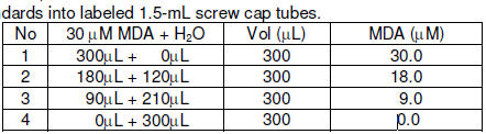
1. Set up standards and samples. Transfer 5 μL Blank, Standard and samples in duplicate wells of a clear bottom 96-well plate.

2. Add 200 μL working reagent and tap lightly to mix.

3. Incubate 30 min at room temperature and read optical density at 510- 630nm (peak absorbance at 590nm).

Procedure using cuvette:

1. Set up test tubes labeled Blank, Standard, Samples. Transfer 20 μL Blank, Standard and samples to appropriately labeled tubes.

2. Add 1000 μL working reagent and tap lightly to mix.

3. Incubate 30 min at room temperature and read optical density at 590nm (510nm-630nm).

CALCULATION

The uric acid concentration of Sample is calculated as

ODBLANK, ODSTANDARD and ODSAMPLE are OD590nm values of Blank, Standard 
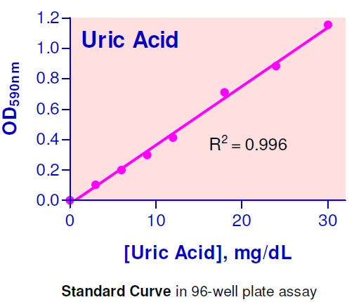
and Sample, respectively. It is not necessary to prepare a calibration curve, because the concentration of the provided standard lies within the linear range. Normal serum uric acid values: 1.0 to 7.0 mg/dL.

Conversions: 1 mg/dL uric acid equals 59.5 μM, 0.001% or 10 ppm.

MATERIALS REQUIRED, BUT NOT PROVIDED

Pipeting devices and accessories (e.g. 5 μL).

Procedure using 96-well plate:

Clear bottom 96-well plates (e.g. Corning Costar). 96-well plate absorbance (590nm) reader.

Procedure using cuvette:

Cuvets for measuring optical density at 510-630nm. Spectrophotometer for measuring absorbance at 590nm.

EXAMPLES:

Samples were assayed using the 96-well protocol. The uric acid content (mg/dL) was 1.3 ± 0.1 (n = 4) for mice serum, 2.6 ± 0.0 (n = 4) for fetal bovine serum (Invitrogen), 1.4 ± 0.1 for goat serum, 1.3 ± 0.1 for rat serum, 2.9 ± 0.1 for rat plasma, 3.4 ± 0.1 for human serum and 1.4 ± 0.1 for human plasma, respectively.

PUBLICATIONS

[1]. Viel, E.C. et al (2008). Xanthine oxidase and mitochondria contribute to vascular superoxide anion generation in DOCA-salt hypertensive rats. Am J Physiol Heart Circ Physiol. 295:H281-H288.

[2]. Kamel, A. H. (2007). Conventional and planar chip sensors for potentiometric assay of uric acid in biological fluids using flow injection analysis. J Pharm Biomed Anal. 45(2):341-348.

[3]. DiSilvestro R. A. et al (2009). Pomegranate extract mouth rinsing effects on saliva measures relevant to gingivitis risk. Phytother Res. 23(8): 1123-1127.