|
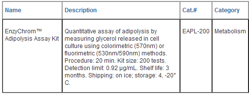
EnzyChromTM
Adipolysis Assay Kit (Cat# EAPL-200)
Quantitative Colorimetric/Fluorimetric
Assay for Adipolysis
DESCRIPTION
Obesity is a chronic condition that
develops from storage of excessive energy in the form of
adipose tissue. The resulting adiposity presents a high
risk factor for diseases such as type 2 diabetes,
cardiovascular diseases, and cancer. ADIPOLYSIS
or lipolysis is a highly regulated process in fat
metabolism, in which triglycerides are broken down into
glycerol and free fatty acids. Rapid, robust and
accurate procedures for adipolysis quantification in
cell culture are very useful in research and drug
discovery. BioAssay Systems' adipolysis assay kit
directly measures glycerol released during adipolysis.
This homogeneous assay uses a single Working Reagent
that combines glycerol kinase, glycerol phosphate
oxidase and color reactions in one step. The color
intensity of the reaction product at 570nm is directly
proportional to glycerol concentration in the sample.
KEY
FEATURES
Sensitive
and accurate.
Use as little as 10 μL samples. Linear detection range
in 96-well plate: 0.92 to 100 μg/mL (10 to 1000 μM)
glycerol for colorimetric assays and 0.2 to 5 μg/mL for
fluorimetric assays.
Rapid
and convenient. The procedure involves addition of a
single working reagent and incubation for 20 min at room
temperature.
Robust
and amenable to HTS assays. Potential interference
by testing drugs is greatly reduced at 570nm. Compatible
with culture media containing phenol red. Assays can be
performed in 96 or 384- well plates.
APPLICATIONS:
Direct
Assays:
adipolysis
(glycerol in cell culture media).
Drug
Discovery/Pharmacology: effects of testing drugs on
adipolysis.
KIT
CONTENTS (bulk reagents available)
Assay
Buffer:
24 mL
Enzyme Mix: 500 μL ATP: 250 μL
Dye
Reagent: 220 μL Standard: 100 μL 100 mM
Glycerol
Storage
conditions. The kit is shipped on dry ice. Store
Assay Buffer at 4°C and other reagents at -20°C. Shelf
life of two months after receipt.
Precautions: reagents are for research use only.
Normal precautions for laboratory reagents should be
exercised while using the reagents. Please refer to
Material Safety Data Sheet for detailed information.
COLORIEMTRIC PROCEDURE
SH-group
containing reagents (e.g. mercaptoethanol, DTT) may
interfere with this assay and should be avoided in
sample preparation. Prior to the assay, equilibrate all
components to room temperature. Keep thawed Enzyme Mix
in a refrigerator or on ice during assays.
1. Cell
Culture. Note:
Cells and
testing drugs are to be provided by the customer and are
not included in this reagent kit.
Grow cells
(e.g. preadipocytes, adipocytes) in culture plate
(24-well, 96-well or 384- well). If desired, treat cells
with testing drugs such as insulin, isoproterenol, and
incubate for the desired time period.
2.
Standards and Samples. Prepare a 100 μg/mL standard
by mixing 10 μL 100 mM glycerol standard with 910 μL in
the same medium used for cell culture. Dilute standard
in the medium as follows. Transfer 10 μL standards into
wells of a clear 96-well assay plate (5 μL for
384-well assay plate).

Collect
cell culture supernatants from culture wells. Such
samples should be assayed immediately or stored at
-20°C. Transfer 10 μL samples (5 μL
for 384-well assay plate)
into separate wells of the assay plate.
3.
Enzyme Reaction. For each assay well, mix 100 μL
Assay Buffer, 2μL Enzyme Mix, 1 μL ATP and 1 μL Dye
Reagent in a clean tube. Transfer
100
μL Working
Reagent into each assay well. Tap plate to mix. For
assays in a 384-well plate, use 50 μL
Working Reagent per well.
4.
Incubate 20 min at room temperature. Read optical
density at 570nm (550-585nm).
Note:
if the Sample OD is higher than the Standard OD at 100
μg/mL,
dilute sample in water and repeat the assay. Multiply
result by the dilution factor.
CALCULATION
Subtract blank OD (#4) from the standard
OD values and plot the OD against standard
concentrations. Determine the slope using linear
regression fitting. The glycerol concentration of Sample
is calculated

ODSAMPLE
and ODMEDIUM
are
optical density values of the sample and medium (#4).
Conversions: 1 μg/mL glycerol equals 10.9 μM.
FLUORIMETRIC PROCEDURE
For
fluorimetric assays, the linear detection range is 0.2
to 5 μg/mL
glycerol. Dilute Standards (#1 to # 4, see
Colorimetric Procedure) as follows: mix 10 μL
standard with 190 μL dH2O. The glycerol
concentrations are now 5.0, 3.0, 1.5 and 0
μg/mL,
respectively.
Cell culture supernatant: dilute by mixing 10
μL cell
culture sample
with 190
μL dH2O
(dilution factor n = 20). Transfer 5 μL of the
diluted standards and samples into separate wells
of a
black
96-well or 384-well plate. Add 50 μL Working Reagent
and tap plate to mix.
Incubate 20 min at room temperature and read
fluorescence at
lex =
530nm and
lem =
585nm.
The glycerol concentration of Sample is calculated as

MATERIALS REQUIRED, BUT NOT PROVIDED
Pipeting devices, centrifuge tubes,
appropriate 96- or 384-well plates and plate reader.

LITERATURE
1. Duncan RE, et al. (2007).
Regulation of lipolysis in adipocytes. Annu Rev Nutr.
27:79-101.
2. Moller F, Roomi MW. (1974). An
enzymatic, spectrophotometric glycerol assay with
increased basic sensitivity. Anal Biochem. 59(1):248-58.
3. MacRae AR. (1977). A
semi-automated enzymatic assay for free glycerol and
triglycerides in serum or plasma. Clin Biochem.
10(1):16-9.
|





















 GENTAUR Germany
Marienbongard 20
GENTAUR Germany
Marienbongard 20
 GENTAUR Italy
 Česká republika
Praha
Česká republika
Praha Danmark
 Finland Helsset
 Ελλάς Αθήνα
 Ireland Dublin
 Luxembourg
 Magyarország
Budapest
Magyarország
Budapest Nederland
 Norge
Oslo
Norge
Oslo Österreich
 Sverige
Stockholm
Sverige
Stockholm Schweiz Züri
 Canada Montreal
 US New York
 日本
日本