|


|
OUR SUPPLIERS
KOMABIOTECH
301,
Gayang Technotown, #1487
Gayang 3 dong,
Gangseo-gu
Seoul 157-793, KOREA
Spherotech, Inc.
27845 Irma Lee Circle, Unit 101
Lake Forest, IL 60045
|
|
Exalpha
Biologicals,
Inc.
2 Shaker Road,
Unit B101
Shirley, MA
01464
SCETI K.K
BIOSCIENCE Export DF Kasumigaseki Place,3-6-7 Kasumigaseki Chiyoda-ku, Tokyo 100-0013 JAPAN
|
EY
Laboratories, Inc. Headquarters
107 N.
Amphlett Blvd
San Mateo, CA. 94401 USA
EXBIO Praha, a.s.
Nad
Safinou II 366
252 42 Vestec
Czech Republic
Sacace Biotechnologies S.r.l.
Via Scalabrini,
44
22100 Como Italy
 GENTAUR BVBA
VAT BE0473327336
Av. de l Armee 68 B4
1040 Brussels
BELGIUM
Tel + 32 16 58 90
45
Fax + 32 16 50 90 45
 GENTAUR France SARL
SIRET 48423788800017
Rue Lagrange, 9
75005 Paris,
France
Tel 01 43 25 01 50
Fax 01 43 25 01 60
 GENTAUR Germany
Marienbongard 20
GENTAUR Germany
Marienbongard 20
52074 Aachen,
Germany
Tel 0241 56 00
99 68
Fax 0241 56 00 47 88
 GENTAUR
Pol Sp. Z.o.o. Ulica
Ogarna 15/19B m2 GENTAUR
Pol Sp. Z.o.o. Ulica
Ogarna 15/19B m2
80-826 GDANSK
Tel 00 48 58 760 77
08
Fax: 00 32 16 50 90
45
 GENTAUR Italy
23015 Milano, Italy
Tel 02 36 00 65
93
Fax 02 36 00 65
94
 Česká republika
Praha
Česká republika
Praha
+420246019719
 Danmark
+4569918806
 Finland Helsset
+358942419041
 Ελλάς Αθήνα
+302111768494
 Ireland Dublin
+35316526556
 Luxembourg
+35220880274
 Magyarország
Budapest
Magyarország
Budapest
+3619980547
 Nederland
+31208080893
 Norge
Oslo Norge
Oslo
+4721031366
 Österreich
+43720880899
 Sverige
Stockholm Sverige
Stockholm
+46852503438
 Schweiz Züri
+41435006251
Northern America
 Canada Montreal
+15149077481
 US New York
+17185132983
Other Countries
0032 (0)16 41 44 07 |
|
|
|
|
|
|

| |
|
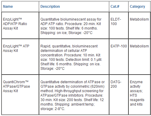
EnzyLightTM
ADP/ATP Ratio Assay Kit
(ELDT-100)
Bioluminescent Assay for ADP/ATP
Ratio
DESCRIPTION
Changes in the ADP/ATP ratio have
been used to differentiate modes of cell death and
viability. Increased levels of ATP and decreased levels
of ADP signify proliferating cells. Conversely,
decreased levels of ATP and increased levels of ADP
represent apoptotic or necrotic cells where the decrease
in ATP and increase in ADP is much more pronounced in
necrosis versus apoptosis. BioAssay Systems’ EnzyLightTM
ADP/ATP Ratio Assay Kit provides a rapid method to
measure ADP and ATP levels for the screening of
apoptosis, necrosis and cell proliferation in mammalian
cells. The assay involves two steps. In the first step,
the working reagent lyses cells to release ATP and ADP.
In the presence of luciferase, ATP immediately
reacts with the Substrate D-luciferin to produce
light. The light intensity is a direct measure of
intracellular ATP concentration.

In the second step, the ADP is
converted to ATP through an enzyme reaction. This newly
formed ATP then reacts with the D-luciferin as in the
first step. Due to a special formulation of the reagent
system which greatly stabilizes the light signal
generated by the luciferase reaction, the luminescence
from the initial ATP measurement remains stable
throughout this assay. Therefore, the second light
intensity measured represents the total ADP and ATP
concentration in the sample. This non-radioactive,
homogeneous cell-based assay is performed in microplates.
The reagent is compatible with all culture media and
with all liquid handling systems for high-throughput
screening applications in 96- well and 384-well plates.
KEY FEATURES
Safe. Non-radioactive assay.
Homogeneous and convenient. "Mix-incubate-measure"
type assay. No wash and reagent transfer steps are
involved.
Robust and amenable to HTS: Z’
factors of 0.5 and above are routinely observed in
96-well and 384-well plates. Can be readily automated on
HTS liquid handling systems for processing thousands of
samples per day.
APPLICATIONS
Apoptosis and Necrosis
determination in cells.
Cell proliferation: effects of
cytokines, growth factor, nutrients.
Drug discovery:
high-throughput screening for anticancer drugs.
KIT CONTENTS
Assay Buffer: 10 mL
Substrate: 120 μL
Cosubstrate 120 μL
ATP Enzyme: 120 μL
ADP Enzyme: 120 μL well. Rock plate
lightly to mix and incubate for desired period of time
(e.g. overnight).
2. ATP Assay. Bring Assay
Buffer, Substrate and Cosubstrate to room temperature.
Thaw enzyme on ice or at 4°C. Fresh Reconstitution is
recommended. Store unused reagents including the enzyme
at -20°C. ATP Reagent. For each 96-well, mix 95
μL Assay Buffer with 1 μL Substrate, 1 μL Cosubstrate
and 1 μL ATP Enzyme. For each 384-well, mix 30 μL Assay
Buffer with 0.3 μL Substrate, 0.3 μL Cosubstrate and 0.3
μL ATP Enzyme. Add ATP Reagent to each well (90
μL/96well, 25 μL/384well) and mix by tapping the plate.
Incubate for 10 minutes at room temperature. Read
luminescence (RLU A) on a luminometer. For most
luminometers (Berthold Luminometer, LJL Analyst, Top
Count, MicroBeta Counters, CLIPR and LeadSeeker),
integration time of 0.1 to 5 sec is appropriate.
3. ADP Assay. Prepare
ADP Reagent: for each 96-well, mix 12 μL dH2O
with 1 μL ADP Enzyme. For each 384-well, mix 3 μL dH2O
with 0.25 μL ADP Enzyme. Add ADP Reagent to each
well (10 μL/96well, 2.5 μL/384well) and mix by tapping
the plate or pipetting up and down. Incubate for 10
minutes at room temperature. Read luminescence (RLU B)
on a luminometer.
4. Calculation of ADP/ATP Ratio.
Subtract RLU A from RLU B, then divide by Data A:

RESULTS INTERPRETATION
The interpretation of different ratios
obtained may vary significantly according to the cell
types and conditions used. However, the following may be
used as guidelines:
1. Test gives markedly elevated ATP
levels with no significant increase in ADP levels in
comparison to control cells = proliferation. (Ratio
<0.1)
2. Test gives lower ATP levels with an
increase in ADP levels in comparison to control cells =
apoptosis. (Ratio ~0.1-1.0)
3. Test gives markedly lower ATP levels
with greatly increased ADP levels in comparison to
control cells = necrosis. (Ratio > 1.0)
LITERATURE
[1]. Bradbury DA, et al (2000).
Measurement of the ADP:ATP ratio in human leukaemic cell
lines can be used as an indicator of cell viability,
necrosis and apoptosis. J Immunol Methods.
240:79-92.
[2]. Chen-Scarabelli C, et al (2004).
Turning necrosis into apoptosis: the exacting task that
can enhance survival. Am Heart J. 148(2):196-9.
[3]. Crouch S, et al (1993). The use of
ATP Bioluminescence as a measure of cell proliferation
and cytotoxicity. J Immunol Methods, 160(1):
81-8.
|
|
|
|
EnzyLightTM
ATP Assay Kit (EATP-100)
Rapid bioluminescent
determination of ATP
DESCRIPTION
Adenosine 5'-triphosphate (ATP) is
the chemical energy for cellular metabolism and is often
referred to as “energy currency" of the cell. ATP is
produced only in living cells during photosynthesis and
cellular respiration and consumed in cellular processes
including biosynthetic reactions, motility and cell
division. It is a key indicator of cellular activity and
has been utilized as a measure of cell viability and
cytotoxicity in research and drug discovery. BioAssay
Systems’ EnzyLightTM ATP Assay Kit provides a rapid
method to measure intracellular ATP. The single working
reagent lyses cells to release ATP, which, in the
presence of luciferase, immediately reacts with
the Substrate D-luciferin to produce light. The
light intensity is a direct measure of intracellular ATP
concentration.

This non-radioactive, homogeneous
cell-based assay is performed in microplates. The
reagent is compatible with all culture media and with
all liquid handling systems for high-throughput
screening applications in 96- well and 384-well plates.
KEY FEATURES
Safe. Non-radioactive assay.
Sensitive and accurate. As low
as 0.1 μM ATP or 40 cells can be quantified.
Homogeneous and convenient.
"Mix-incubate-measure" type assay. No wash and reagent
transfer steps are involved.
Robust and amenable to HTS: Z’
factors of > 0.5 are routinely observed in 96-well and
384-well plates. Can be readily automated on HTS liquid
handling systems for processing thousands of samples per
day.
APPLICATIONS
ATP determination in cells and
other biological samples.
KIT CONTENTS
Assay Buffer: 10 mL
Substrate: 120 μL
ATP Enzyme: 1 20 μL
Standard: 100 μL 3 mM ATP
Storage conditions: store all
reagents at -20°C. This kit is shipped on dry ice. Shelf
life of at least 6 months.
Precautions: reagents are for
research use only. Normal precautions for laboratory
reagents should be exercised while using the reagents.
Please refer to Material Safety Data Sheet for detailed
information.
ASSAY PROCEDURE
1. Standard Curve. Prepare
1000 μL 30 μM ATP Premix by mixing 10 μL 3 mM Standard
and 990 μL distilled water (for cell culture samples
dilute ATP in culture media). Dilute standard as
follows. Transfer 100 μL standards into wells of a white
opaque 96-well plate.

Samples.
Add 100 μL sample per well in separate wells. For tissue
samples, homogenize 20 mg sample in 200
μL of cold
phosphate-buffered saline, spin at 12,000 g for 5 min to
pellet any debris. Transfer 1-100
μL
supernatant to each well and bring the volume to 100
μL
with PBS. Test several doses of the sample and choose
the readings that are within the standard curve range
for ATP calculation. For cell cultures, plate cells (100
μL/96well plate, 25 μL/384well
plate) in white opaque tissue culture plates. If
desired, add 5
μL
test compounds and controls dissolved in PBS or culture
medium per well. Rock plate lightly to mix and incubate
for desired period of time (e.g. overnight).
2.
Assay. Bring Assay Buffer and Substrate to room
temperature. Thaw enzyme on ice or at 4°C. Fresh
Reconstitution is recommended. Store unused reagents
including the enzyme at -20°C. For each 96-well, mix 95
μL Assay Buffer with 1 μL
Substrate and 1 μL ATP Enzyme. Add 90
μL
Reconstituted Reagent to each well. For each 384-well,
mix 30
μL Assay
Buffer with 0.3 μL
Substrate and 0.3
μL ATP
Enzyme. Add 25 μL
Reconstituted Reagent to each well. Mix by tapping the
plate. Incubate for 10 minutes at room temperature. 3.
Read luminescence on a luminometer. For most
luminometers (Berthold Luminometer, LJL Analyst, Top
Count, MicroBeta Counters, CLIPR and LeadSeeker),
integration time of 0.1 to 5 sec is appropriate.
GENERAL CONSIDERATIONS
Signal
stability.
After adding the Reconstituted Reagent, the luminescence
signal is stable for about 20 min and decreases slow
thereafter.

LITERATURE
[1].
Kangas L, et al. (1984). Bioluminescence of cellular ATP:
a new method for evaluating agents in vitro. Medical
Biology, 62: 338 - 343.
[2].
Zhelev Z, et al (2004). Phenothiazines suppress
proliferation and induce apoptosis in cultured leukemic
cells without any influence on the viability of normal
lymphocytes. Phenothiazines and leukemia.
Cancer
Chemother Pharmacol.
53(3):267-75.
[3].
Ingram PR, et al (2004). A comparison of the effects of
ocular preservatives on mammalian and microbial ATP and
glutathione levels. Free Radic Res.
|
|
|
|
QuantiChromTM ATPase/GTPase Assay Kit
(DATG-200)
DESCRIPTION
ATPases
and GTPases catalyze the decomposition of ATP or GTP
into ADP or GDP and free phosphate ion. These enzymes
play key roles in transport, signal transduction,
protein biosynthesis and cell differentiation. BioAssay
Systems’ QuantiChromTM ATPase/GTPase Assay Kit offers a
highly sensitive method for determining ATPase/GTPase
activities in a microplate format. Its proprietary
formulation features a single reagent for accurate
determination of enzyme activity in 30 min at room
temperature. The improved malachite green reagent forms
a stable dark green color with liberated phosphate,
which is measured on a plate reader (600 - 660 nm).
KEY
FEATURES
High
sensitivity:
detection of as little of 2 pmoles of free phosphate.
Fast
and convenient: single reagent, homogeneous
“mix-and-measure” assay allows quantitation of enzyme
activity within 30 minutes.
Robust
and amenable to HTS: detection at 620nm greatly
reduces potential interference by colored compounds. Z’
factors of >0.7 are observed in 96-well and 384-well
plates. Can be readily automated on HTS liquid handling
systems.
APPLICATIONS
Determination
of ATPase
and GTPase activity.
Drug
Discovery: high-throughput screen for ATPase or
GTPase inhibitors.
KIT
CONTENTS: 200 ASSAYS IN 96-WELL PLATE
Reagent:
50 mL Assay Buffer: 10 mL
Standard: 1mL 1 mM phosphate
Storage
conditions. The reagents and standard are stable for
one year when stored at 4°C.
Precautions: reagent contains 0.27 M H2SO4. Normal
precautions for laboratory reagents should be exercised
while using the reagents. Please refer to Material
Safety Data Sheet for detailed information.
Important: All reagents must
be brought to room temperature before use. Before each
assay, it is important to check that enzyme preparations
and assay buffers do not contain free phosphate. This
can be conveniently done by adding 200 μL of the Reagent
to 40 μL sample solution. The blank OD values at 620 nm
should be lower than 0.3. If the OD readings are higher
than 0.3, check phosphate level. Lab detergents may
contain high levels of phosphate. Make sure that lab
wares are free from contaminating phosphate after
thorough washes.
ACTIVITY DETERMINATION IN 96-WELL PLATE
1.
Preparation of phosphate standards. Prepare 800 μL
Premix solution containing 50 μM phosphate by mixing 40
μL 1 mM phosphate standard with 760 μL distilled water.
Number the tubes. Dilute standards as shown in the
following Table. Pipette 40 μL standard in duplicate
into wells of a clear-bottom 96-well plate.

2.
Perform a series dilution of enzyme in assay buffer.
Set up 40-μL reactions and a control with no enzyme in
separate wells. Incubate the reaction for desired period
of time (e.g. 30 min).

3. Add
200 μL Reagent and incubate 30 min at room
temperature. Please note: use of a multi-chanel pipettor
is recommended. The Reagent terminates the enzyme
reaction and generates color with the free phosphate
produced in the enzyme reaction.
4. Read
OD620nm on a plate reader.
5.
Enzyme activity. Calculate DOD values by subtracting
OD values in reaction and control wells. Choose an
enzyme concentration that gives a DOD of 0.5 to 1, this
will ensure that substrate hydrolysis (<10%) is within
the linear kinetics of reaction. Compute the
concentration of free phosphate produced from the
standard curve. 1 Unit of activity is the amount of
enzyme that catalyzes the production of 1 μmole of free
phosphate per minute under the assay conditions.
INHIBITOR ASSAY IN 96-WELL PLATE
To
evaluate an inhibitor or perform HTS, use the optimal
enzyme concentration determined above. Incubate enzyme
and inhibitor first for a certain period of time, before
adding the substrate. At the end of reaction, add 200 μL
Reagent for phosphate determination.

ASSAYS IN 384-WELL PLATE
The
procedure is similar as in the 96-well plate assay,
except that 20 μL standards or 20 μL reaction mixture
(10 μL Assay Buffer, 5 μL 4 mM ATP, 5 μL enzyme) are
mixed with 80 μL Reagent.
GENERAL CONSIDERATIONS
Materials. Use ultrapure ATP and GTP. The provided
2x assay buffer contains 40 mM Tris, 80 mM NaCl, 8 mM
MgAc2, 1 mM EDTA, pH 7.5. Other buffers (Hepes, Mes,
Mops) can be used. Assay is compatible with 1 mM DTT,
2mM b-mercaptoethanol, 0.5 mg/mL BSA and 5% DMSO.

Phosphate assays in 96-well plate.
1. H2O, 2. Phosphate, 3. ATP in H2O, 4. ATP/Phosphate in
H2O, 5, 7, 9, 11, 13: ATP in Assay Buffer with, where
indicated, 0.5 mg/mL BSA, 1 mM DTT, 2 mM
b-mercaptoethanol (bME)
and 5% DMSO. 6, 8, 10, 12, 14: ATP/Phosphate in Assay
Buffer. Phosphate and ATP were at 50
μM
and 1 mM, respectively. The assay is not affected by
these components.
|
|
|
|




















 日本
日本