|
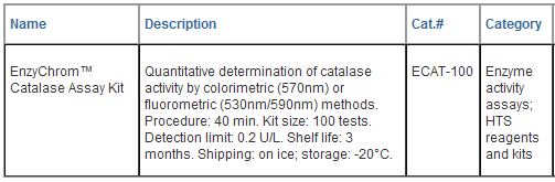
EnzyChromTM Catalase Assay Kit
(ECAT-100)
Quantitative Colorimetric/Fluoriemtric
Catalase Determination
DESCRIPTION
CATALASE (EC 1.11.1.6), is an
ubiquitous antioxidant enzyme that catalyzes the
decomposition of hydrogen peroxide (H2O2) to water and
oxygen.

By
preventing excessive H2O2 build up, catalase allows
important cellular processes which produce H2O2 as a
byproduct to occur safely. Deficiency in catalase
activity has been associated with grey hair and
peroxisomal disorder acatalasia. Simple, direct and
high-throughput assays for catalase activity find wide
applications. BioAssay Systems' improved assay directly
measures catalase degradation of H2O2 using a redox dye.
The change in color intensity at 570nm or fluorescence
intensity (lem/ex = 585/530nm) is directly proportional
to the catalase activity in the sample.
KEY
FEATURES
Sensitive
and accurate.
Use 10 μL sample. Linear detection range 0.2 to 5 U/L
catalase activity.
Simple
and Convenient. The procedure involves adding a
Substrate to sample, incubation for 30 min, followed by
a Detection Reagent and reading the optical density or
fluorescence intensity.
APPLICATIONS:
Direct
Assays:
catalase
activity in biological samples e.g. serum, plasma,
urine, saliva, cell culture etc.
Drug
Discovery/Pharmacology: effects of drugs on catalase
activity.
KIT
CONTENTS:
Assay
Buffer:
25 mL
HRP Enzyme: 120 μL Dye Reagent: 120 μL
H2O2
Solution: 100 μL 3% H2O2 Positive Control: 8
μL Catalase
Storage
conditions. The kit is shipped on ice. Store all
components at -20°C. Shelf life of three months after
receipt.
Precautions: reagents are for research use only.
Normal precautions for laboratory reagents should be
exercised while using the reagents. Please refer to
Material Safety Data Sheet for detailed information.
SAMPLE PREPARATION
Tissue (10
mg) and cells (106) are homogenized in 200 μL cold Assay
Buffer. Centrifuge 10 min at 14,000 rpm to pellet any
debris. Use clear supernatant for assay.
Note:
SH-containing reagents (e.g. b–mercaptoethanol,
dithiothreitol) are known to interfere in this assay and
should be kept below 10
μM
in the sample.
ASSAY
PROCEDURE
1.
Reagent Preparation. Equilibrate all components to
room temperature. Briefly centrifuge all tubes before
opening. Keep thawed HRP Enzyme on ice. For colorimetric
assays, use a clear flat- bottom 96-well plate. For
fluorimetric assays, use a solid black flat-bottom
96-well plate. Samples and Controls: transfer 10
μL sample into wells of the 96-well plate. In addition,
for each assay run, prepare one sample blank well that
contains only 10 μL Assay Buffer. Add 400 μL Assay
Buffer to Positive Control tube and mix well. Transfer
10 μL of the reconstituted Positive Control into
separate wells.
Note:
(1). For unknown samples, perform several dilutions to
ensure that catalase activity is within the linear range
0.2 to 5 U/L. (2) The provided catalase serves as a
positive control to ensure assay is working and should
not be used to calculate the Sample catalase activity.
2.
Enzyme Reaction. Mix 5 μL 3% H2O2 and 914 μL dH2O
(final 4.8 mM). Prepare enough 50 μM H2O2 Substrate for
sample, positive control and sample blank by mixing, for
each well, 1 μL of the 4.8 mM H2O2 with 95 μL Assay
Buffer.
Note:
diluted H2O2 is not stable. Prepare fresh dilutions for
each experiment.
Add 90 μL
of the 50 μM Substrate to these wells to initiate the
catalase reaction. Tap plate quick to mix. Incubate 30
min at room temperature. During the incubation time,
proceed with Steps 3 and 4 below.
3. H2O2
Standard Curve. Mix 40μL of the 4.8 mM H2O2 with
440 μL dH2O to yield 400 μM H2O2. Prepare standards as
shown in the Table below. Transfer 10 μL standards into
separate wells of the 96-well plate. Add 90 μL Assay
Buffer to the standards.

4.
Detection. Prepare enough Detection Reagent by
mixing, for each reaction well (Sample, Control and
Standard wells), 102 μL Assay Buffer, 1 μL Dye Reagent
and 1 μL HRP Enzyme. At the end of the 30 min incubation
(Step 2), add 100 μL Detection Reagent per well.
Tap plate to mix. Incubate for 10 min.
5. Read
optical density at 570nm (550 to 585nm) or fluorescence
intensity at lem/ex = 585/530nm.
CALCULATION
Subtract
blank value (#4) from the standard values and plot the
DOD or DF against standard concentrations. Determine the
slope and calculate the catalase activity of Sample,

RSAMPLE
Blank
and RSAMPLE
are
optical density or fluorescence intensity readings of
the Sample Blank and Sample, respectively. Slope is
determined from the standard curve. 30 min is the
catalase reaction time. n is the sample dilution
factor. Unit definition: one unit is the amount
of catalase that decomposes 1 μmole of H2O2 per min at
pH 7.0 and room temperature.
MATERIALS REQUIRED, BUT NOT PROVIDED
Pipetting
devices, centrifuge tubes, uncoated 96-well plates,
optical density plate reader, fluorescence plate reader,
homogenizer etc.

LITERATURE
1. Cowell, D.C. et al (1994). The rapid
potentiometric detection of catalase positive
microorganisms. Biosens Bioelectron. 9(2):131-138.
2. Góth, L. (1991). A simple method for
determination of serum catalase activity and revision of
reference range. Clin Chim Acta. 196:143-151.
3. Kurasaki, M. et al (1986). Increased
erythrocyte catalase activity in patients with
hyperthyroidism. Horm Metab Res. 18:56-59.
|





















 GENTAUR Germany
Marienbongard 20
GENTAUR Germany
Marienbongard 20
 GENTAUR Italy
 Česká republika
Praha
Česká republika
Praha Danmark
 Finland Helsset
 Ελλάς Αθήνα
 Ireland Dublin
 Luxembourg
 Magyarország
Budapest
Magyarország
Budapest Nederland
 Norge
Oslo
Norge
Oslo Österreich
 Sverige
Stockholm
Sverige
Stockholm Schweiz Züri
 Canada Montreal
 US New York
 日本
日本