Home ] Up ] Immunology Infectious agents Cellular Biology Molecular biology Instruments Chimical Product PCR ELISA Sites Komabiotech products Product NEW antibodies NEW Cells Culturs EPIGENTEK Electrophoresis Contact Us EnoGene SACACE Albumin—OsrHSA GENTAUR NEWS Antibodies & Supporting ToolsHome ] Up ] Luciferase Electrophoresis  Albumin—OsrHSA Pricelist:  http://www.diagrade.com/search.php  

Google

       Pricelist 2010 

Home
Up

OUR SUPPLIERS

KOMABIOTECH
301, Gayang Technotown, #1487 Gayang 3 dong, Gangseo-gu
Seoul 157-793, KOREA

Spherotech, Inc.
27845 Irma Lee Circle, Unit 101
Lake Forest, IL 60045

Exalpha Biologicals, Inc.
2 Shaker Road, Unit B101
Shirley, MA 01464

SCETI K.K
BIOSCIENCE
Export                                   DF Kasumigaseki Place,3-6-7 Kasumigaseki Chiyoda-ku, Tokyo 100-0013 JAPAN 

EY Laboratories, Inc. Headquarters
107 N. Amphlett Blvd
San Mateo, CA. 94401 USA

EXBIO Praha, a.s.
Nad Safinou II 366
252 42  Vestec
Czech Republic


Sacace Biotechnologies S.r.l.

Via Scalabrini, 44
22100 Como Italy


redcoon België GENTAUR BVBA

VAT BE0473327336

Av. de l Armee 68 B4

1040 Brussels BELGIUM

  Tel + 32 16 58 90 45 

Fax + 32 16 50 90 45

GENTAUR France SARL

SIRET 48423788800017

Rue Lagrange, 9

75005 Paris, France

 Tel 01 43 25 01 50

Fax 01 43 25 01 60 

GENTAUR Germany Marienbongard 20

52074 Aachen, Germany

Tel  0241 56 00 99 68                    Fax 0241 56 00 47 88 

GENTAUR Pol Sp. Z.o.o. Ulica Ogarna 15/19B m2

80-826 GDANSK

Tel 00 48 58 760 77 08

Fax: 00 32 16 50 90 45

GENTAUR Italy

23015 Milano, Italy

 Tel 02 36 00 65 93

Fax 02 36 00 65 94

Česká republika Praha
+420246019719
 

Danmark

+4569918806 

Finland Helsset
+358942419041

Ελλάς Αθήνα
+302111768494
 

Ireland Dublin
+35316526556
 

Luxembourg
+35220880274
 

Magyarország Budapest
+3619980547
 

Nederland
+31208080893
 

Norge Oslo
+4721031366
 

Österreich
+43720880899
 

Sverige Stockholm
+46852503438
 

Schweiz Züri
+41435006251


Northern America 

Canada Montreal
+15149077481
 

US New York
+17185132983


Other Countries redcoon België
0032 (0)16 41 44 07


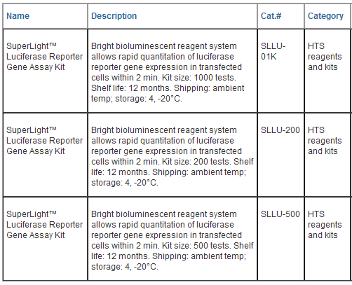
SuperLightTM Luciferase Reporter Gene Assay Kits

Bioluminescent Assay for Promoter Regulated Luciferase Expression

DESCRIPTION

The SuperLightTM Luciferase Reporter Gene Assay is based on the quantitation of luciferase expression in mammalian, yeast or E. coil cells, using luciferin and ATP as substrates. The reaction results in light production which can be conveniently measured on a luminometer.

This bioluminescent reporter gene assay is extremely sensitive and is especially suitable for quantifying luciferase expression in recombinant cells. This ultra-sensitive, homogeneous cell-based assay only requires adding a single reagent to the cells and measuring the light intensity after a short incubation step (2 minutes). Assays can be performed in tubes, cuvettes or multi-well plates. All kit components are compatible with culture media and with all liquid handling systems. With an extended luminescence emission kinetics (half-life 40 min), the SuperLightTM luciferase assays are especially suitable for high-throughput screening in 96-well, 384-well and 1536-well plates. In addition, the reagent provided in the kits has been formulated for maximum sensitivity, reproducibility and long shelf-life. Applications for this kit include gene regulation studies and high-throughput screening of gene modulators.

KEY FEATURES

High sensitivity and wide detection range: detection of as little of 2 fg luciferase and as few as 4 cells. Plus, the emitted light is linear over seven orders of magnitude.

Compatible with routine laboratory and HTS formats: assays can be performed in tubes or microplates, on LJL Analyst, Berthold Luminometer, Top-Count, MicroBeta counters, chemiluminescent image plate readers (CLIPR/LeadSeeker). Assay reagents compatible with all liquid handling systems.

Fast and convenient: homogeneous “mix-and-measure” assay allows detection of luciferase levels within 10 minutes. The optimally combined reagent system allows a single addition step, and simultaneous cell lysis and detection.

Robust and amenable to HTS: Z’ factors of 0.6 to 0.8 are observed in 96- well and 384-well plates. Can be readily automated on HTS liquid handling systems.

APPLICATIONS

Gene Regulation: gene expression level, characterization of promoter activity, modulation of gene expression by receptors, transcription factors and small molecules.

Drug Discovery: high-throughput screen for gene modulators.

Storage conditions. Store the Reagent in the provided amber tube at - 20°C and the Assay Buffer at 2-8°C. Shelf life: 12 month.

Precautions: reagents are for research use only. Normal precautions for laboratory reagents should be exercised while using the reagents. Please refer to Material Safety Data Sheet for detailed information.

PROCEDURES

The SuperLightTM Luciferase Reporter Gene Assay is based on the bioluminescence generated during the luciferin/luciferase reaction. The reconstituted reagent has been optimized to combine cell lysis and detection into one single step. Phenol red in culture media does not interfere in this assay. All data in the Technical Notes were obtained in media containing phenol red.

Important: fresh reconstitution of the Reagent in Assay Buffer is recommended, although the reconstituted Reagent may be stable for up to 4 weeks when stored at -20 °C.

Procedure using 96-well plate:

1. Plate and culture cells (80 μL) in white opaque 96-well tissue culture plates. Typical culture medium contains DMEM, 10% fetal bovine serum and antibiotics (penicillin/ streptomycin, gentamycin, etc). Amino acids and other nutrients can be added to the culture medium. Assays can be performed on either adherent cells or cells in suspension. The cells can be either stably or transiently transfected with the luciferase gene. Culture volume can vary from 50 to 100 μL, although 80 μL is used in this protocol. Blank control wells containing no cells should also be prepared.

2. Add test compounds and controls to cells. Mix well and incubate for the cells desired period of time. Incubation time for gene regulation studies can be from several hours up to 3 days. It is recommended that assays be run in duplicate or triplicate. A volume of 20 μL compounds in PBS or culture medium is recommended.

3. Reconstitute the Reagent. First equilibrate the Reagent and Assay Buffer to room temperature. Then simply combine the Assay Buffer and Reagent by pipetting a small volume (e.g. 1 mL) buffer to the Reagent tube. Vortex briefly and pipet the reconstituted solution to the Assay Buffer bottle. Repeat this step to transfer all Reagent to the Assay Buffer bottle. Mix by inversion until the Reagent is thoroughly dissolved. After this is done, mark the bottle label as Reconstituted Reagent.

4. Add 100 μL (per 80 μL of cell culture) of the reconstituted Reagent to each well and mix well with the cells. Incubate for 2 minutes at room temperature. The volume of the reagent can be adjusted depending on the volume of cell culture.

5. Measure luminescence on a luminometer. The integration time can be 1 sec to 2 min depending on the luciferase expression level and instrument sensitivity. For most luminometers (Berthold Luminometer, LJL Analyst, Top Count, MicroBeta Counters, CLIPR and LeadSeeker), integration 1 to 5 sec is appropriate.

Procedure using 384-well plate:

1. Plate and culture cells (25 μL) in white opaque 384-well tissue culture plates. Culture volume can vary from 20 to 50 μL, although 25 μL is used in this protocol. Set up blank control wells containing no cells.

2. Add test compounds and controls to cells. Mix well and incubate for the cells desired period of time. A volume of 5 μL compounds in PBS or culture medium is recommended.

3. Reconstitute the Reagent using the same procedure as for the 96-well assay.

4. Add 30 μL (per 25 μL of cell culture) of the reconstituted Reagent per well and mix well with the cells. Incubate for 2 minutes at room temperature. The volume of the reagent can be adjusted depending on the volume of cell culture.

5. Measure luminescence on a luminometer. The integration time can be 1 sec to 2 min depending on the luciferase expression level and instrument sensitivity. For most luminometers (Berthold Luminometer, LJL Analyst, Top Count, MicroBeta Counters, CLIPR and LeadSeeker), integration 1 to 5 sec is appropriate.

GENERAL CONSIDERATIONS

Incubation time. Both the luciferin/luciferase reaction and cell lysis are fast, so incubation for 2 to 10 minutes following reagent addition is generally enough for mammalian cells (e.g. HEK293, CHO).

Cell number. The optimized reporter gene assay reagent is very sensitive to luciferase (detection limit 2 fg) and exhibits linearity over seven orders of magnitude. As few as 4 cells can be determined and a linear response is still observed with as many as 80,000 cells per 96- well. For assay optimization, it is recommended that the optimal number of cells per well be determined by serial dilution of cells. Cells can be adherent or in suspension cultures.

Cell lysis and mixing. For the sake of convenience, the addition of 1 volume of reconstituted reagent to 1 volume of cells allows a sufficient mixing. No additional mixing is required since the specially formulated buffer instantly lyses mammalian cells.

DATA ANALYSIS

The light intensity (RLU) is directly proportional to the luciferase concentration. For dose-response studies, the data are plotted against compound concentration and the EC50 for gene up-regulator compound and IC50 for a gene down regulator compound can be determined by non-linear regression analysis using Prism or other data analysis tools.

PUBLICATIONS

1. Zhao, L. and Haslam, D.B. (2005). A quantitative and highly sensitive luciferase-based assay for bacterial toxins that inhibit protein synthesis. J Med Microbiol 54:1023–1030.

2. Saenz JB, Doggett TA, and Haslam (2007). Identification and characterization of small molecules that inhibit intracellular toxin transport. Infection and Immunity 75(9): 4552–4561.

3. Gentry, M. et al. (2007). Role of primary human alveolar epithelial cells in host defense against Francisella tularensis Infection. Infection and Immunity 75(8): 3969-3978.

4. Michael, K. et al. (2007). Microplate orbital mixing improves highthroughput cell-based reporter assay readout. J Biomol Screen 12(1):140-144.

TECHNICAL NOTES

The SuperLightTM Luciferase Reporter Gene Assay Kit has been specially optimized and formulated to provide a sensitive, convenient and robust assay for gene expression and regulation studies in mammalian cells.  Keyfeatures of the kits are as follows:

High sensitivity and wide detection range: detection of as little of 2 fg luciferase and as few as 4 cells. Plus, the emitted light is linear over seven orders of magnitude.

Compatible with routine laboratory and HTS formats: assays can be performed in tubes or microplates, on LJL Analyst, Berthold Luminometer, Top-Count, MicroBeta counters, chemiluminescent image plate readers (CLIPR/LeadSeeker). Assay reagents compatible with all liquid handling systems.

Fast and convenient: homogeneous “mix-and-measure” assay allows detection of luciferase levels within 10 minutes. The optimally combined reagent system allows a single addition step and simultaneous cell lysis and detection.

Robust and amenable to HTS. Z’ factors of 0.7 to 0.9 are routinely observed in 96-well and 384-well plates. Can be readily automated on HTS liquid handling systems.


SuperLightTM Luciferase Reporter Gene Assay Kits

Bioluminescent Assay for Promoter Regulated Luciferase Expression

DESCRIPTION

The SuperLightTM Luciferase Reporter Gene Assay is based on the quantitation of luciferase expression in mammalian, yeast or E. coil cells, using luciferin and ATP as substrates. The reaction results in light production which can be conveniently measured on a luminometer.

This bioluminescent reporter gene assay is extremely sensitive and is especially suitable for quantifying luciferase expression in recombinant cells. This ultra-sensitive, homogeneous cell-based assay only requires adding a single reagent to the cells and measuring the light intensity after a short incubation step (2 minutes). Assays can be performed in tubes, cuvettes or multi-well plates. All kit components are compatible with culture media and with all liquid handling systems. With an extended luminescence emission kinetics (half-life 40 min), the SuperLightTM luciferase assays are especially suitable for high-throughput screening in 96-well, 384-well and 1536-well plates. In addition, the reagent provided in the kits has been formulated for maximum sensitivity, reproducibility and long shelf-life. Applications for this kit include gene regulation studies and high-throughput screening of gene modulators.

KEY FEATURES

High sensitivity and wide detection range: detection of as little of 2 fg luciferase and as few as 4 cells. Plus, the emitted light is linear over seven orders of magnitude.

Compatible with routine laboratory and HTS formats: assays can be performed in tubes or microplates, on LJL Analyst, Berthold Luminometer, Top-Count, MicroBeta counters, chemiluminescent image plate readers (CLIPR/LeadSeeker). Assay reagents compatible with all liquid handling systems.

Fast and convenient: homogeneous “mix-and-measure” assay allows detection of luciferase levels within 10 minutes. The optimally combined reagent system allows a single addition step, and simultaneous cell lysis and detection.

Robust and amenable to HTS: Z’ factors of 0.6 to 0.8 are observed in 96- well and 384-well plates. Can be readily automated on HTS liquid handling systems.

APPLICATIONS

Gene Regulation: gene expression level, characterization of promoter activity, modulation of gene expression by receptors, transcription factors and small molecules.

Drug Discovery: high-throughput screen for gene modulators.

Storage conditions. Store the Reagent in the provided amber tube at - 20°C and the Assay Buffer at 2-8°C. Shelf life: 12 month.

Precautions: reagents are for research use only. Normal precautions for laboratory reagents should be exercised while using the reagents. Please refer to Material Safety Data Sheet for detailed information.

PROCEDURES

The SuperLightTM Luciferase Reporter Gene Assay is based on the bioluminescence generated during the luciferin/luciferase reaction. The reconstituted reagent has been optimized to combine cell lysis and detection into one single step. Phenol red in culture media does not interfere in this assay. All data in the Technical Notes were obtained in media containing phenol red.

Important: fresh reconstitution of the Reagent in Assay Buffer is recommended, although the reconstituted Reagent may be stable for up to 4 weeks when stored at -20 °C.

Procedure using 96-well plate:

1. Plate and culture cells (80 μL) in white opaque 96-well tissue culture plates. Typical culture medium contains DMEM, 10% fetal bovine serum and antibiotics (penicillin/ streptomycin, gentamycin, etc). Amino acids and other nutrients can be added to the culture medium. Assays can be performed on either adherent cells or cells in suspension. The cells can be either stably or transiently transfected with the luciferase gene. Culture volume can vary from 50 to 100 μL, although 80 μL is used in this protocol. Blank control wells containing no cells should also be prepared.

2. Add test compounds and controls to cells. Mix well and incubate for the cells desired period of time. Incubation time for gene regulation studies can be from several hours up to 3 days. It is recommended that assays be run in duplicate or triplicate. A volume of 20 μL compounds in PBS or culture medium is recommended.

3. Reconstitute the Reagent. First equilibrate the Reagent and Assay Buffer to room temperature. Then simply combine the Assay Buffer and Reagent by pipetting a small volume (e.g. 1 mL) buffer to the Reagent tube. Vortex briefly and pipet the reconstituted solution to the Assay Buffer bottle. Repeat this step to transfer all Reagent to the Assay Buffer bottle. Mix by inversion until the Reagent is thoroughly dissolved. After this is done, mark the bottle label as Reconstituted Reagent.

4. Add 100 μL (per 80 μL of cell culture) of the reconstituted Reagent to each well and mix well with the cells. Incubate for 2 minutes at room temperature. The volume of the reagent can be adjusted depending on the volume of cell culture.

5. Measure luminescence on a luminometer. The integration time can be 1 sec to 2 min depending on the luciferase expression level and instrument sensitivity. For most luminometers (Berthold Luminometer, LJL Analyst, Top Count, MicroBeta Counters, CLIPR and LeadSeeker), integration 1 to 5 sec is appropriate.

Procedure using 384-well plate:

1. Plate and culture cells (25 μL) in white opaque 384-well tissue culture plates. Culture volume can vary from 20 to 50 μL, although 25 μL is used in this protocol. Set up blank control wells containing no cells.

2. Add test compounds and controls to cells. Mix well and incubate for the cells desired period of time. A volume of 5 μL compounds in PBS or culture medium is recommended.

3. Reconstitute the Reagent using the same procedure as for the 96-well assay.

4. Add 30 μL (per 25 μL of cell culture) of the reconstituted Reagent per well and mix well with the cells. Incubate for 2 minutes at room temperature. The volume of the reagent can be adjusted depending on the volume of cell culture.

5. Measure luminescence on a luminometer. The integration time can be 1 sec to 2 min depending on the luciferase expression level and instrument sensitivity. For most luminometers (Berthold Luminometer, LJL Analyst, Top Count, MicroBeta Counters, CLIPR and LeadSeeker), integration 1 to 5 sec is appropriate.

GENERAL CONSIDERATIONS

Incubation time. Both the luciferin/luciferase reaction and cell lysis are fast, so incubation for 2 to 10 minutes following reagent addition is generally enough for mammalian cells (e.g. HEK293, CHO).

Cell number. The optimized reporter gene assay reagent is very sensitive to luciferase (detection limit 2 fg) and exhibits linearity over seven orders of magnitude. As few as 4 cells can be determined and a linear response is still observed with as many as 80,000 cells per 96- well. For assay optimization, it is recommended that the optimal number of cells per well be determined by serial dilution of cells. Cells can be adherent or in suspension cultures.

Cell lysis and mixing. For the sake of convenience, the addition of 1 volume of reconstituted reagent to 1 volume of cells allows a sufficient mixing. No additional mixing is required since the specially formulated buffer instantly lyses mammalian cells.

DATA ANALYSIS

The light intensity (RLU) is directly proportional to the luciferase concentration. For dose-response studies, the data are plotted against compound concentration and the EC50 for gene up-regulator compound and IC50 for a gene down regulator compound can be determined by non-linear regression analysis using Prism or other data analysis tools.

PUBLICATIONS

1. Zhao, L. and Haslam, D.B. (2005). A quantitative and highly sensitive luciferase-based assay for bacterial toxins that inhibit protein synthesis. J Med Microbiol 54:1023–1030.

2. Saenz JB, Doggett TA, and Haslam (2007). Identification and characterization of small molecules that inhibit intracellular toxin transport. Infection and Immunity 75(9): 4552–4561.

3. Gentry, M. et al. (2007). Role of primary human alveolar epithelial cells in host defense against Francisella tularensis Infection. Infection and Immunity 75(8): 3969-3978.

4. Michael, K. et al. (2007). Microplate orbital mixing improves highthroughput cell-based reporter assay readout. J Biomol Screen 12(1):140-144.

TECHNICAL NOTES

The SuperLightTM Luciferase Reporter Gene Assay Kit has been specially optimized and formulated to provide a sensitive, convenient and robust assay for gene expression and regulation studies in mammalian cells.  Keyfeatures of the kits are as follows:

High sensitivity and wide detection range: detection of as little of 2 fg luciferase and as few as 4 cells. Plus, the emitted light is linear over seven orders of magnitude.

Compatible with routine laboratory and HTS formats: assays can be performed in tubes or microplates, on LJL Analyst, Berthold Luminometer, Top-Count, MicroBeta counters, chemiluminescent image plate readers (CLIPR/LeadSeeker). Assay reagents compatible with all liquid handling systems.

Fast and convenient: homogeneous “mix-and-measure” assay allows detection of luciferase levels within 10 minutes. The optimally combined reagent system allows a single addition step and simultaneous cell lysis and detection.

Robust and amenable to HTS. Z’ factors of 0.7 to 0.9 are routinely observed in 96-well and 384-well plates. Can be readily automated on HTS liquid handling systems.


SuperLightTM Luciferase Reporter Gene Assay Kits

Bioluminescent Assay for Promoter Regulated Luciferase Expression

DESCRIPTION

The SuperLightTM Luciferase Reporter Gene Assay is based on the quantitation of luciferase expression in mammalian, yeast or E. coil cells, using luciferin and ATP as substrates. The reaction results in light production which can be conveniently measured on a luminometer.

This bioluminescent reporter gene assay is extremely sensitive and is especially suitable for quantifying luciferase expression in recombinant cells. This ultra-sensitive, homogeneous cell-based assay only requires adding a single reagent to the cells and measuring the light intensity after a short incubation step (2 minutes). Assays can be performed in tubes, cuvettes or multi-well plates. All kit components are compatible with culture media and with all liquid handling systems. With an extended luminescence emission kinetics (half-life 40 min), the SuperLightTM luciferase assays are especially suitable for high-throughput screening in 96-well, 384-well and 1536-well plates. In addition, the reagent provided in the kits has been formulated for maximum sensitivity, reproducibility and long shelf-life. Applications for this kit include gene regulation studies and high-throughput screening of gene modulators.

KEY FEATURES

High sensitivity and wide detection range: detection of as little of 2 fg luciferase and as few as 4 cells. Plus, the emitted light is linear over seven orders of magnitude.

Compatible with routine laboratory and HTS formats: assays can be performed in tubes or microplates, on LJL Analyst, Berthold Luminometer, Top-Count, MicroBeta counters, chemiluminescent image plate readers (CLIPR/LeadSeeker). Assay reagents compatible with all liquid handling systems.

Fast and convenient: homogeneous “mix-and-measure” assay allows detection of luciferase levels within 10 minutes. The optimally combined reagent system allows a single addition step, and simultaneous cell lysis and detection.

Robust and amenable to HTS: Z’ factors of 0.6 to 0.8 are observed in 96- well and 384-well plates. Can be readily automated on HTS liquid handling systems.

APPLICATIONS

Gene Regulation: gene expression level, characterization of promoter activity, modulation of gene expression by receptors, transcription factors and small molecules.

Drug Discovery: high-throughput screen for gene modulators.

Storage conditions. Store the Reagent in the provided amber tube at - 20°C and the Assay Buffer at 2-8°C. Shelf life: 12 month.

Precautions: reagents are for research use only. Normal precautions for laboratory reagents should be exercised while using the reagents. Please refer to Material Safety Data Sheet for detailed information.

PROCEDURES

The SuperLightTM Luciferase Reporter Gene Assay is based on the bioluminescence generated during the luciferin/luciferase reaction. The reconstituted reagent has been optimized to combine cell lysis and detection into one single step. Phenol red in culture media does not interfere in this assay. All data in the Technical Notes were obtained in media containing phenol red.

Important: fresh reconstitution of the Reagent in Assay Buffer is recommended, although the reconstituted Reagent may be stable for up to 4 weeks when stored at -20 °C.

Procedure using 96-well plate:

1. Plate and culture cells (80 μL) in white opaque 96-well tissue culture plates. Typical culture medium contains DMEM, 10% fetal bovine serum and antibiotics (penicillin/ streptomycin, gentamycin, etc). Amino acids and other nutrients can be added to the culture medium. Assays can be performed on either adherent cells or cells in suspension. The cells can be either stably or transiently transfected with the luciferase gene. Culture volume can vary from 50 to 100 μL, although 80 μL is used in this protocol. Blank control wells containing no cells should also be prepared.

2. Add test compounds and controls to cells. Mix well and incubate for the cells desired period of time. Incubation time for gene regulation studies can be from several hours up to 3 days. It is recommended that assays be run in duplicate or triplicate. A volume of 20 μL compounds in PBS or culture medium is recommended.

3. Reconstitute the Reagent. First equilibrate the Reagent and Assay Buffer to room temperature. Then simply combine the Assay Buffer and Reagent by pipetting a small volume (e.g. 1 mL) buffer to the Reagent tube. Vortex briefly and pipet the reconstituted solution to the Assay Buffer bottle. Repeat this step to transfer all Reagent to the Assay Buffer bottle. Mix by inversion until the Reagent is thoroughly dissolved. After this is done, mark the bottle label as Reconstituted Reagent.

4. Add 100 μL (per 80 μL of cell culture) of the reconstituted Reagent to each well and mix well with the cells. Incubate for 2 minutes at room temperature. The volume of the reagent can be adjusted depending on the volume of cell culture.

5. Measure luminescence on a luminometer. The integration time can be 1 sec to 2 min depending on the luciferase expression level and instrument sensitivity. For most luminometers (Berthold Luminometer, LJL Analyst, Top Count, MicroBeta Counters, CLIPR and LeadSeeker), integration 1 to 5 sec is appropriate.

Procedure using 384-well plate:

1. Plate and culture cells (25 μL) in white opaque 384-well tissue culture plates. Culture volume can vary from 20 to 50 μL, although 25 μL is used in this protocol. Set up blank control wells containing no cells.

2. Add test compounds and controls to cells. Mix well and incubate for the cells desired period of time. A volume of 5 μL compounds in PBS or culture medium is recommended.

3. Reconstitute the Reagent using the same procedure as for the 96-well assay.

4. Add 30 μL (per 25 μL of cell culture) of the reconstituted Reagent per well and mix well with the cells. Incubate for 2 minutes at room temperature. The volume of the reagent can be adjusted depending on the volume of cell culture.

5. Measure luminescence on a luminometer. The integration time can be 1 sec to 2 min depending on the luciferase expression level and instrument sensitivity. For most luminometers (Berthold Luminometer, LJL Analyst, Top Count, MicroBeta Counters, CLIPR and LeadSeeker), integration 1 to 5 sec is appropriate.

GENERAL CONSIDERATIONS

Incubation time. Both the luciferin/luciferase reaction and cell lysis are fast, so incubation for 2 to 10 minutes following reagent addition is generally enough for mammalian cells (e.g. HEK293, CHO).

Cell number. The optimized reporter gene assay reagent is very sensitive to luciferase (detection limit 2 fg) and exhibits linearity over seven orders of magnitude. As few as 4 cells can be determined and a linear response is still observed with as many as 80,000 cells per 96- well. For assay optimization, it is recommended that the optimal number of cells per well be determined by serial dilution of cells. Cells can be adherent or in suspension cultures.

Cell lysis and mixing. For the sake of convenience, the addition of 1 volume of reconstituted reagent to 1 volume of cells allows a sufficient mixing. No additional mixing is required since the specially formulated buffer instantly lyses mammalian cells.

DATA ANALYSIS

The light intensity (RLU) is directly proportional to the luciferase concentration. For dose-response studies, the data are plotted against compound concentration and the EC50 for gene up-regulator compound and IC50 for a gene down regulator compound can be determined by non-linear regression analysis using Prism or other data analysis tools.

PUBLICATIONS

1. Zhao, L. and Haslam, D.B. (2005). A quantitative and highly sensitive luciferase-based assay for bacterial toxins that inhibit protein synthesis. J Med Microbiol 54:1023–1030.

2. Saenz JB, Doggett TA, and Haslam (2007). Identification and characterization of small molecules that inhibit intracellular toxin transport. Infection and Immunity 75(9): 4552–4561.

3. Gentry, M. et al. (2007). Role of primary human alveolar epithelial cells in host defense against Francisella tularensis Infection. Infection and Immunity 75(8): 3969-3978.

4. Michael, K. et al. (2007). Microplate orbital mixing improves highthroughput cell-based reporter assay readout. J Biomol Screen 12(1):140-144.

TECHNICAL NOTES

The SuperLightTM Luciferase Reporter Gene Assay Kit has been specially optimized and formulated to provide a sensitive, convenient and robust assay for gene expression and regulation studies in mammalian cells.  Keyfeatures of the kits are as follows:

High sensitivity and wide detection range: detection of as little of 2 fg luciferase and as few as 4 cells. Plus, the emitted light is linear over seven orders of magnitude.

Compatible with routine laboratory and HTS formats: assays can be performed in tubes or microplates, on LJL Analyst, Berthold Luminometer, Top-Count, MicroBeta counters, chemiluminescent image plate readers (CLIPR/LeadSeeker). Assay reagents compatible with all liquid handling systems.

Fast and convenient: homogeneous “mix-and-measure” assay allows detection of luciferase levels within 10 minutes. The optimally combined reagent system allows a single addition step and simultaneous cell lysis and detection.

Robust and amenable to HTS. Z’ factors of 0.7 to 0.9 are routinely observed in 96-well and 384-well plates. Can be readily automated on HTS liquid handling systems.