|


|
OUR SUPPLIERS
KOMABIOTECH
301,
Gayang Technotown, #1487
Gayang 3 dong,
Gangseo-gu
Seoul 157-793, KOREA
Spherotech, Inc.
27845 Irma Lee Circle, Unit 101
Lake Forest, IL 60045
|
|
Exalpha
Biologicals,
Inc.
2 Shaker Road,
Unit B101
Shirley, MA
01464
SCETI K.K
BIOSCIENCE Export DF Kasumigaseki Place,3-6-7 Kasumigaseki Chiyoda-ku, Tokyo 100-0013 JAPAN
|
EY
Laboratories, Inc. Headquarters
107 N.
Amphlett Blvd
San Mateo, CA. 94401 USA
EXBIO Praha, a.s.
Nad
Safinou II 366
252 42 Vestec
Czech Republic
Sacace Biotechnologies S.r.l.
Via Scalabrini,
44
22100 Como Italy
 GENTAUR BVBA
VAT BE0473327336
Av. de l Armee 68 B4
1040 Brussels
BELGIUM
Tel + 32 16 58 90
45
Fax + 32 16 50 90 45
 GENTAUR France SARL
SIRET 48423788800017
Rue Lagrange, 9
75005 Paris,
France
Tel 01 43 25 01 50
Fax 01 43 25 01 60
 GENTAUR Germany
Marienbongard 20
GENTAUR Germany
Marienbongard 20
52074 Aachen,
Germany
Tel 0241 56 00
99 68
Fax 0241 56 00 47 88
 GENTAUR
Pol Sp. Z.o.o. Ulica
Ogarna 15/19B m2 GENTAUR
Pol Sp. Z.o.o. Ulica
Ogarna 15/19B m2
80-826 GDANSK
Tel 00 48 58 760 77
08
Fax: 00 32 16 50 90
45
 GENTAUR Italy
23015 Milano, Italy
Tel 02 36 00 65
93
Fax 02 36 00 65
94
 Česká republika
Praha
Česká republika
Praha
+420246019719
 Danmark
+4569918806
 Finland Helsset
+358942419041
 Ελλάς Αθήνα
+302111768494
 Ireland Dublin
+35316526556
 Luxembourg
+35220880274
 Magyarország
Budapest
Magyarország
Budapest
+3619980547
 Nederland
+31208080893
 Norge
Oslo Norge
Oslo
+4721031366
 Österreich
+43720880899
 Sverige
Stockholm Sverige
Stockholm
+46852503438
 Schweiz Züri
+41435006251
Northern America
 Canada Montreal
+15149077481
 US New York
+17185132983
Other Countries
0032 (0)16 41 44 07 |
|
|
|
|
|
|

| |
|
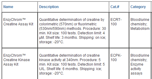
EnzyChromTM Creatine Assay Kit (ECRT-100)
Quantitative Colorimetric/Fluorimetric
Determination of Creatine
DESCRIPTION
CREATINE
is present in
vertebrates and helps to supply energy to muscle.
In humans and
animals, approximately half of creatine originates from food (mainly
from fresh meat). Creatine supplementation has been investigated as a
possible therapeutic approach for the treatment of muscular,
neuromuscular, neurological and neurodegenerative diseases. Simple,
direct and automation-ready procedures for measuring creatine are
popular in research and drug discovery. BioAssay Systems' creatine assay
is based on enzymatic reactions leading to formation of a pink colored
product. The optical density at 570 nm or fluorescence intensity at
lem/ex
= 590/530 nm is directly proportional to the creatine concentration in
the sample.
KEY FEATURES
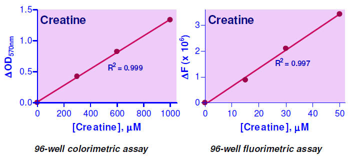
High sensitivity and wide linear range .
Use 10 μL
sample. Linear detection range 4 to 1000
μM (colorimetric) or 0.5 to 50 μM (fluorimetric).
Homogeneous and simple procedure .
Simple “mix-and-measure” procedure allows reliable quantitation of
creatine within 30 minutes.
APPLICATIONS
Direct Assays:
creatine in
biological samples (e.g. serum, plasma, urine, saliva etc).
KIT CONTENTS (100 tests in 96-well plates)
Assay Buffer : 20 mL
Enzyme A: 120 μL
Enzyme B: 220 μL Standard: 400 μL 20 mM
creatine
Dye Reagent: 220 μL
Storage conditions .
The kit is shipped chilled. Store all components at -20°C. Shelf life of
at least 3 months.
Precautions :
reagents are for research use only. Normal precautions for laboratory
reagents should be exercised while using the reagents. Please refer to
Material Safety Data Sheet for detailed information.
PROCEDURES
Sample preparation :
SH-group containing reagents (e.g. mercaptoethanol, DTT) and EDTA may
interfere with this assay and should be avoided in sample preparation.
Solid samples can be extracted by homogenization in distilled water
(dH2O) and filtered or centrifuged. Liquid samples (e.g. serum, plasma
and urine) can be assayed directly.
Colorimetric Procedure
1. Standards and Samples .
Equilibrate all components to room temperature. Briefly centrifuge tubes
before opening. Prepare a 1000
μM
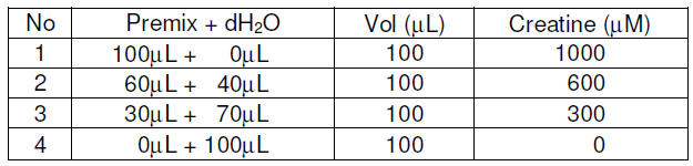
creatine Standard Premix by mixing 15 μL
of the 20 mM Standard and 285 μL dH2O.
Dilute Standard as follows.
Transfer 10 μ L
standards into separate wells of a clear, flat-bottom 96-
well plate.
Transfer 10
μL
of each sample into two separate wells, one serving as a
sample blank
well (RBLANK) and one as a sample well (RSAMPLE).
2.
Enzyme Reaction.
For each standard and sample well, prepare Working Reagent by mixing 90
μL
Assay Buffer, 1 μL
Enzyme A, 1 μL
Enzyme B
and 1 μL
Dye Reagent. Add 90 μL
Working Reagent to the
four Standards
and the Sample Wells.
Prepare blank control reagent by mixing 90 μ L
Assay Buffer, 1 μL
Enzyme B and 1 μL
Dye Reagent (i.e. no Enzyme A). Add 90 μL
Blank control reagent only to the Sample
Blank Wells.
Tap plate to mix. Incubate 30 min at room temperature.
3. Read OD570nm.
Fluorimetric Procedure
The fluorimetric procedure is the same as for the
colorimetric assay, except that (1) the detection range is up to 50 μ M
creatine and (2) a black, flat-bottom 96-well plate is used. Creatine
standards of 0, 15, 30 and 50 μM
are prepared. After incubation for 30 min at room temperature, read
fluorescence intensity at lex
= 530 nm and lem
= 590 nm.
CALCULATION
Subtract the standard values from the blank value (#4)
and plot the DOD or DF against standard concentrations. Determine the
slope and calculate the creatine concentration of Sample,

RSAMPLE and RBLANK are optical density or fluorescence
intensity readings of the Sample and Sample Blank, respectively. n
is the sample dilution factor.
Note: if the calculated creatine concentration is
higher than 1000 μM in the colorimetric assay or 50 μM in the
fluorimetric assay, dilute sample in dH2O and repeat assay. Multiply
result by the dilution factor n.
Conversions: 1000 μM creatine equals 13.1 mg/dL
or 131 ppm.
MATERIALS REQUIRED, BUT NOT PROVIDED
Pipeting devices, and clear flat-bottom 96-well plates
and optical density plate reader for colorimetric assays; black
flat-bottom 96-well plate and fluorescence intensity plate reader for
fluorimetric assays.
LITERATURE
1. Jabs, C.M. et al (1988). Plasma creatine
determination using a luminescence method. Biochem Med Metab Biol.
39(3):267-272.
2. Anderson, D.R. et al (1957). Determination of
creatine in biological fluids. Biochem J. 67(2): 258-262.
3. Delanghe, J. et al (1986). Early diagnosis of acute
myocardial infarction by enzymatic urinary creatine determination. Clin
Chem. 32(8):1611. |
|
EnzyChromTM Creatine Kinase Assay Kit (ECPK-100)
Colorimetric Determination of Creatine Kinase Activity
at 340 nm
DESCRIPTION
CREATINE KINASE
(CK), also known
as creatine phosphokinase
(CPK), is an
enzyme (EC 2.7.3.2) expressed predominantly in skeletal muscle, smooth
muscle and the brain. The CK enzyme consists of two subunits, which can
be either B (brain type) or M (muscle type), and hence three different
isoenzymes: CK-MM, CK-BB and CK-MB. CK catalyzes the conversion of
creatine to phosphocreatine, consuming adenosine triphosphate (ATP) and
generating adenosine diphosphate (ADP) and the reverse reaction. CK is
often determined routinely in emergency patients with chest pain and
acute renal failure. Elevation of CK is an indication of damage to
muscle and has been associated with injury, rhabdomyolysis, myocardial
infarction, myositis, myocarditis, malignant hyperthermia and
neuroleptic malignant syndrome, etc. Lower levels can be an indication
of alcoholic liver disease and rheumatoid arthritis. Simple, direct and
automation-ready procedures for measuring CK activity are very
desirable. BioAssay Systems' QuantiChromTM Creatine Kinase Assay Kit is
based on enzyme coupled reactions in which creatine phosphate and ADP is
converted to creatine and ATP by CK, the generated ATP is used to
phosphorylate glucose by hexokinase to generate glucose-6-phosphate,
which is then oxidized by NADP in the presence of glucose-6- phosphate
dehydrogenase.
The produced NADPH, measured at 340 nm, is proportionate
to the CK activity in the sample.
APPLICATIONS
Direct Assays: CK in serum, plasma and other biological
samples.
Pharmacology: effects of drugs on CK activity.
KEY FEATURES
Sensitive and accurate .
Detection range: 5 to 300 U/L creatine kinase in 96-well plate assay.
Convenient .
The procedure involves adding a single working reagent, and reading the
optical density at 10 min and 40 min at room temperature or 37°C.
High-throughput .
Can be readily automated as a high-throughput 96- well plate assay for
thousands of samples per day.
KIT CONTENTS (100 tests in 96-well plates)
Assay Buffer: 12 mL Substrate Solution: 1.0 mL
Enzyme Mix: 120 μL Calibrator: 150 μ L
Storage conditions .
This kit is shipped on dry ice. Store all reagents at -20°C. Shelf life
of at least 6 months (see expiry dates on labels).
Precautions :
reagents are for research use only. Normal precautions for laboratory
reagents should be exercised while using the reagents. Please refer to
Material Safety Data Sheet for detailed information.
PROCEDURES
Sample Preparation .
Use non-hemolyzed samples. Samples should be assayed within 4 hours of
blood collection if they remain at room temperature, or within 12 hours
if stored at 4°C. Samples can be stored at -80°C and should be thawed
only once. If turbidity is observed, centrifuge sample and use clear
supernatant for assay.
Reagent Reconstitution .
Bring all components to room temperature.
For each reaction well, mix 10
μL Substrate Solution, 100 μL
Assay Buffer and 1 μL
Enzyme Mix. Fresh reconstitution is recommended. If the assay is to be
carried out at 37°C, warm up the Reconstituted Reagent at 37°C.
1.
Calibrator: transfer 110 μL water and (10 μL Calibrator + 100 μL
water) into separate wells of a clear bottom 96-well plate.
Samples: transfer 10 μL samples into separate wells.
Add 100 μ L
Reconstituted Reagent and tap plate to mix.
2. Reaction. Incubate at room temperature or
37° C.
CK is fully
activated within
10 min by glutathione provided in the Substrate
Solution. Read OD340nm at 10 min and again at 40 min.
3.
Calculation. Calculate sample CK activity using the equation,

OD40min and OD10min are OD340nm values at 40 min and 10
min for the sample. ODCALIBRATOR and ODH2O are OD340nm values of the
Calibrator and water blank at 40 min. The value 100 is the equivalent
activity (U/L) of the Calibrator under the assay conditions. Unit
definition: one unit of CK will transfer 1 μ mole
of phosphate from phosphocreatine to ADP per min at pH 6.0.
Note: If the CK activity is
expected to be higher than 300 U/L, read OD340nm at 10 min and again at
15 min. To calculate the CK activity replace (OD40min – OD10min) with
(OD15min – OD10min) and replace the factor 100 with 600 in the above
equation. Linear range: 30 to 1,800 U/L CK activity.
MATERIALS REQUIRED, BUT NOT PROVIDED
Pipeting (multi-channel) devices. Clear-bottom
96-well plates (e.g. Corning Costar) and plate reader.
GENERAL CONSIDERATIONS
This assay is based on an enzyme-catalyzed kinetic
reaction. Addition of Reconstituted Reagent should be quick and mixing
should be brief but thorough. Use of multi-channel pipettor is
recommended.
EXAMPLES
Samples were assayed in duplicate using the 96-well
protocol. The CK activity (U/L) was 12.0 ± 0.9 for a rat serum sample,
11.0 ± 0.5 for human serum, 28 ± 1 for human plasma, 9.0 ± 0.8 for mouse
serum and 49 ± 2 for bovine serum.

PUBLICATIONS
1. Itaka K et al (2010). Polyplex nanomicelle promotes
hydrodynamic gene introduction to skeletal muscle. J Control Release.
143(1):112-9.
2. Steen C et al (2010). Reduced Creatine Kinase B
Activity in Multiple Sclerosis Normal Appearing White Matter. PLoS One
5(5):e10811.
3. Kwon S et al. (2010). ASB9 interacts with ubiquitous
mitochondrial creatine kinase and inhibits mitochondrial function. BMC
Biol. 8:23. |
|




















 日本
日本