



















Spherotech, Inc.
27845 Irma Lee Circle, Unit 101
Lake Forest, IL 60045
27845 Irma Lee Circle, Unit 101
Lake Forest, IL 60045
|
|
|
|
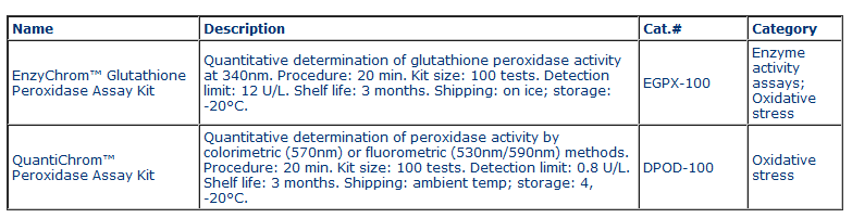
EnzyChromTM Glutathione Peroxidase Assay Kit (EGPX-100) Quantitative Colorimetric Glutathione Peroxidase Determination DESCRIPTION GLUTATHIONE PEROXIDASE (GPX, EC 1.11.1.9) represents an enzyme family with peroxidase activity whose main biological role is to protect the organism from oxidative damage. It helps prevent lipid peroxidation of cellular membranes by removing free peroxide in the cell. GPX catalyzes the following reaction with glutathione reductase (GR), GPX 2 GSH + H2O2 GS-SG + 2 H2O, GR GS-SG + NADPH 2 GSH + NADP+ Simple, direct and high-throughput assays for GPX activity find wide applications. BioAssay Systems' improved assay directly measures NADPH consumption in the enzyme coupled reactions. The measured decrease in optical density at 340nm is directly proportional to the enzyme activity in the sample. KEY FEATURES Sensitive and accurate. Use 10 μL sample. Linear detection range 12 to 300 U/L GPX activity. APPLICATIONS Direct Assays: GPX activity in biological samples. Drug Discovery/Pharmacology: effects of drugs on GPX activity. KIT CONTENTS Assay Buffer: 25 mL GR Enzyme: 1 mL Glutathione: 240 μL NADPH: 40 μL H2O2 Solution: 100 μL 3% H2O2 Positive Control: 9 μL Glutathione Peroxidase (GPX) Storage conditions. The kit is shipped on ice. Store all components at -20 °C. Shelf life of three months after receipt. Precautions: reagents are for research use only. Normal precautions for laboratory reagents should be exercised while using the reagents. Please refer to Material Safety Data Sheet for detailed information. SAMPLE PREPARATION All samples should be clear and free of any turbidity or particles. Liquid samples (e.g. non-hemolyzed serum, plasma) can be assayed directly. Homogenize tissue (10 mg) and cells (106) in 200 μL cold 1 x PBS and then centrifuge 10 min at 14,000 rpm to pellet any debris. Use the clear supernatant for the assay. If not assayed immediately, freeze supernatant at -80°C (stable for 1 month). ASSAY PROCEDURE 1. Reagent Preparation. Equilibrate all components to room temperature. Briefly centrifuge all tubes before opening. Add 360 μL dH2O to the NADPH tube (final 35 mM). Add 500 μL Assay Buffer to the “Positive Control” tube. Vortex tubes to mix. Keep these reconstituted reagent tubes on ice. Unused reagents are stable for three weeks when stored frozen at -20°C. 2. NADPH Standards and Samples. Mix 45 μL of the reconstituted 35 mM NADPH with 217 μL dH2O (final 6 mM). Dilute standards as shown in the Table below. Transfer 10 μL standards into wells of a clear flatbottom 96-well plate. Add 190 μL Assay Buffer to all standard wells.
Transfer 10 μL sample and 10 μL reconstituted GPX Positive Control into separate wells of the 96-well plate. In addition, for each assay run, include a background control that only contains 10 μL Assay Buffer. Note: (1). For unknown samples, perform several dilutions to ensure that GPX activity is within the linear range of 12 to 300 U/L. (2) The provided GPX serves as a positive control to ensure assay is working and should not be used to calculate the Sample GPX activity. 3. Assay. Prepare enough Working Reagent for Sample and Control wells by mixing, for each well, 85 μL Assay Buffer, 2 μL Glutathione, 2 μL 35 mM NADPH and 8 μL GR enzyme. Add 90 μL Working Reagent quickly to the Sample/Control wells. Tap plate to mix. Dilute 8 μL 3% H2O2 with 1992 μL dH2O (final 3.5 mM). Prepare enough 0.35 mM H2O2 Reagent by mixing, for each Sample/Control well, 12 μL 3.5 mM with 108 μL dH2O. Use this Reagent within one hour. With a multi-channel pipettor, add 100 μL 0.35 mM H2O2 Reagent to all Sample and Control wells. Tap plate quickly to mix well contents thoroughly. Immediately read OD340nm (time zero, OD0) and again at 4 min (OD4). Note: if calculated GPX activity is higher than 300 U/L, or initial OD340nm is >1.5 in sample wells, dilute sample in dH2O and repeat assay. Multiply the results by the dilution factor. CALCULATION Use OD values at 4 min for NADPH standards. Subtract blank value (#4) from the standard values. Plot the DOD against standard concentrations and determine the slope of the standard curve. Calculate the DODs = (OD0 – OD4) for the samples and DODB = (OD0 – OD4) for the background control. Calculate the GPX activity of Sample,
The factor 1000 converts mmoles to μmoles. n is the sample dilution factor. Unit definition: one unit is the amount of GPX that produces 1 μmole of GS-SG per min at pH 7.6 and room temperature. MATERIALS REQUIRED, BUT NOT PROVIDED Pipetting devices, centrifuge tubes, clear flat-bottom uncoated 96-well plates, plate reader capable of reading optical density at 340nm every minute, homogenizer (e.g. Sigma # Z359971) etc.
LITERATURE 1. Paglia, D.E. and Valentine, W.N. (1967). Studies on the quantitative and qualitative characterization of erythrocyte glutathione peroxidase. J Lab Clin Med. 70:158-169. 2. Jacobson, B. et al. (1988). Adaptation of glutathione peroxidase assay to the Technicon RA-1000. Clin Chem. 34:2164-2165. 3. Pascual, P. et al. (1992). Direct assay of glutathione peroxidase activity using high-performance capillary electrophoresis. J Chromatogr. 581:49-56. QuantiChromTM Peroxidase Assay Kit (Cat# DPOD-100) Quantitative Colorimetric/Fluorimetric Peroxidase Determinations DESCRIPTION PEROXIDASES (EC number 1.11.1.x) catalyze the following oxidationreduction reactions:
For many peroxidases the optimal substrate is hydrogen peroxide (H2O2), but others are more active with organic hydroperoxides such as lipid peroxides. In the cell, peroxidases destroy toxic hydroxide radicals that are formed as byproducts during aerobic respiration. The peroxidases represent a large family of enzymes that are found in animals (e.g. myeloperoxidase-like enzymes), plant, fungi and bacteria (cytochrome-c peroxidase like enzymes such a horseradish peroxidase). Simple, direct and automation-ready procedures for determining peroxidase activity find wide applications. BioAssay Systems' peroxidase assay uses H2O2 and an electron donor dye that forms a pink color during the peroxidase reaction. The optical density (570nm) or fluorescence intensity (lexc = 530nm, lem = 590nm) is a direct measure of the enzyme activity. KEY FEATURES Use as little as 10 μL samples. Linear detection range: colorimetric assays 4 to 1000 IU/L, fluorimetric assays 0.8 to 25 IU/L peroxidase. KIT CONTENTS Assay Buffer (pH 7.0): 20 mL Dye Reagent: 60 μL Stabilized H2O2: 100 μL 3% S top Reagent: 12 mL Calibrator: 5 mL (equivalent to 1000 IU/L) Storage conditions. The kit is shipped on blue ice. Store Assay Buffer and Dye Reagent at -20°C, all other reagents at 4°C. Shelf life of three months after receipt. Precautions: reagents are for research use only. Normal precautions for laboratory reagents should be exercised while using the reagents. Please refer to Material Safety Data Sheet for detailed information. PROCEDURES Sample Preparation. Samples can be prepared according to established methods [1-3]. It is prudent to test multiple sample dilutions to ensure activity is in the linear range. Reagent Preparation: Bring all reagents to room temperature prior to assay. Dilute 3% H2O2 in Assay Buffer to 0.6% and use within one hour. 96-Well Colorimetric Assay Procedure. Use clear flat-bottom plates for colorimetric assays 1. Transfer 200 μL H2O and 200 μL Calibrator into two wells of a clear flat-bottom 96-well plate. Transfer 10 μL H2O (sample blank), 10 μL sample to separate wells. Prepare fresh Working Reagent for each reaction well by mixing 95 μL Assay Buffer, 0.5 μL Dye Reagent and 0.5 μL freshly diluted 0.6% H2O2. Add 90 μL Working Reagent to each sample well. Tap plate to mix and incubate for 10 min at room temperature. 2. Add 100 μL Stop Reagent to the sample blank and sample wells. Tap plate to mix and read OD570nm. Note: if Sample OD values are higher than that of the Calibrator, dilute sample in Assay Buffer, repeat assay and multiply results by the dilution factor. Peroxidase activity is calculated from the OD values of the sample, sample blank, calibrator and H2O wells.
Unit definition: one unit of enzyme will catalyze the oxidization by H2O2 of 1 μmole dye reagent per min under the assay conditions. 96-Well Fluorimetric Assay Procedure. Use black flat-bottom plates. If desirable, a peroxidase standard (e.g. Sigma Aldrich Cat# P6140 horseradish peroxidase) can be run together with the samples. 1. Transfer 10 μL H2O, 10 μL sample to separate wells. Prepare fresh Working Reagent for each sample well by mixing 95 μL Assay Buffer, 0.5 μL Dye Reagent and 0.5 μL freshly diluted 0.6% H2O2. Add 90 μL Working Reagent to each well. Tap plate to mix and incubate for 10 min at room temperature. 2. Add 100 μL Stop Reagent to the sample blank and sample wells. Tap plate to mix and read fluorescence at 590nm (lexc= 530nm). 384-Well Fluorimetric Assay Procedure. Use 5 μL H2O, 5 μL sample, 35 μL Working Reagent prepared from 38 μL Assay Buffer, 0.2 μL Dye Reagent, 0.2 μL 0.6% H2O2. Use 40 μL Stop Reagent. GENERAL CONSIDERATIONS This assay is based on a kinetic reaction, the use of a multi-channel pipettor for adding the Working Reagent and Stop Reagent is recommended. MATERIALS REQUIRED, BUT NOT PROVIDED Pipeting devices, centrifuge tubes, clear flat bottom 96/384-well plates for colorimetric assays, black 96/384-well plate for fluorimetric assays and plate reader.
LITERATURE 1. Kokkinakis DM, Brooks JL. (1979). Tomato Peroxidase: Purification, Characterization, and Catalytic Properties. Plant Physiol. 63(1):93-99. 2. Pettigrew GW, Seilman S. (1982). Purification and properties of a cross-linked complex between cytochrome c and cytochrome c peroxidase. Biochem J. 201(1):9-18. 3. Smith AL, et al. (1974). Brain polymorphonuclear leukocyte quantitation by peroxidase assay. Infect Immun. 10(2):356-60. | ||||||||||||
|
|


 GENTAUR Germany
Marienbongard 20
GENTAUR Germany
Marienbongard 20
 GENTAUR Italy
 Česká republika
Praha
Česká republika
Praha Danmark
 Finland Helsset
 Ελλάς Αθήνα
 Ireland Dublin
 Luxembourg
 Magyarország
Budapest
Magyarország
Budapest Nederland
 Norge
Oslo
Norge
Oslo Österreich
 Sverige
Stockholm
Sverige
Stockholm Schweiz Züri
 Canada Montreal
 US New York
 日本
日本